Skip to Content
Merck
All Photos(1)

Documents

06-1036

Sigma-Aldrich

Anti-March5 Antibody

serum, from rabbit

Synonym(s):

membrane-associated ring finger (C3HC4) 5, Membrane-associated RING finger protein 5, RING finger protein 153, E3 ubiquitin-protein ligase MARCH5, Mitochondrial ubiquitin ligase, Membrane-associated RING-CH protein V

Sign Into View Organizational & Contract Pricing


About This Item

UNSPSC Code:
12352203
eCl@ss:
32160702
NACRES:
NA.41

biological source

rabbit

Quality Level

antibody form

serum

antibody product type

primary antibodies

clone

polyclonal

species reactivity

human

species reactivity (predicted by homology)

mouse (based on 100% sequence homology), rat (based on 100% sequence homology)

technique(s)

immunocytochemistry: suitable
western blot: suitable

NCBI accession no.

UniProt accession no.

shipped in

dry ice

target post-translational modification

unmodified

Gene Information

human ... MARCHF5(54708)

General description

Membrane-associated RING-CH protein 5 (March5), also known as NKP66, is thought to be a mitochondrial ubiquitin ligase that is localized on the mitochondrial membrane. March5 has a Plant Homeo-Domain (PHD) motif that is essential for E3 ubiquitin ligase action. MARCH 5 is also thought to have four transmembrane domains. March5 has been associated with mitochondrial fission and division. March5 also seems to have a defensive role against mitochondrial dysfunction by interacting with mutant superoxide dismutase 1 (mSOD1) and preventing mSOD1 from causing damage. mSOD1 damage has been linked to the onset and progression of amyotrophic lateral sclerosis.

Immunogen

KLH-conjugated linear peptide corresponding to human March5.
.

Application

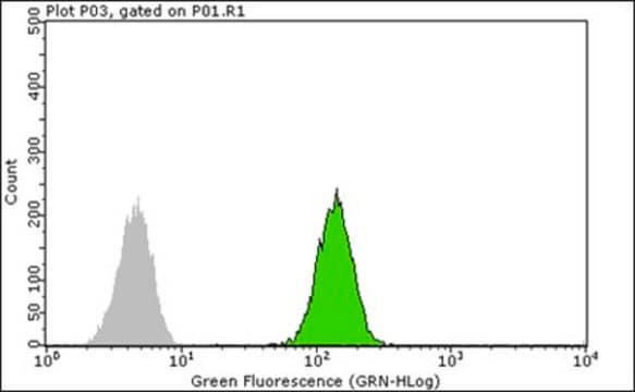
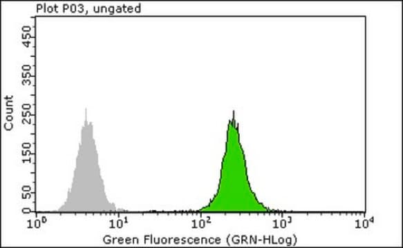
Anti-March5 Antibody is a Rabbit Polyclonal Antibody for detection of March5 also known as Membrane-associated RING finger protein 5 & has been validated in WB, ICC.
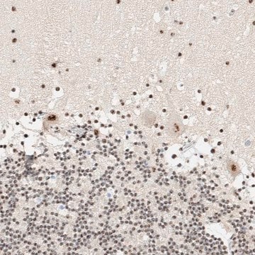
Immunocytochemistry Analysis: A representative lot of this antibody was used by an independent laboratory in IC. (Korfali, N., et al. (2010). Mol Cell Proteomics. 9:2571-2585.)
Research Category
Epigenetics & Nuclear Function
Research Sub Category
Ubiquitin & Ubiquitin Metabolism

Quality

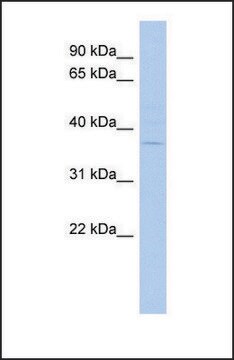
Evaluated by Western Blot in HL-60 cell lysate.

Western Blot Analysis: A 1:2,000 dilution of this antibody detected March5 on 10 µg of HL-60 cell lysate.

Target description

~52 kDa observed

Physical form

Rabbit polyclonal serum containing 0.05% sodium azide.
Unpurified

Storage and Stability

Stable for 1 year at -20°C from date of receipt.
Handling Recommendations: Upon receipt and prior to removing the cap, centrifuge the vial and gently mix the solution. Aliquot into microcentrifuge tubes and store at -20°C. Avoid repeated freeze/thaw cycles, which may damage IgG and affect product performance.

Analysis Note

Control
HL-60 cell lysate

Disclaimer

Unless otherwise stated in our catalog or other company documentation accompanying the product(s), our products are intended for research use only and are not to be used for any other purpose, which includes but is not limited to, unauthorized commercial uses, in vitro diagnostic uses, ex vivo or in vivo therapeutic uses or any type of consumption or application to humans or animals.

Not finding the right product?  

Try our Product Selector Tool.

Storage Class Code

10 - Combustible liquids

WGK

WGK 1


Certificates of Analysis (COA)

Search for Certificates of Analysis (COA) by entering the products Lot/Batch Number. Lot and Batch Numbers can be found on a product’s label following the words ‘Lot’ or ‘Batch’.

Already Own This Product?

Find documentation for the products that you have recently purchased in the Document Library.

Visit the Document Library

The leukocyte nuclear envelope proteome varies with cell activation and contains novel transmembrane proteins that affect genome architecture.
Korfali, N; Wilkie, GS; Swanson, SK; Srsen, V; Batrakou, DG; Fairley, EA; Malik et al.
Molecular and Cellular Proteomics null
Manuel D Haschka et al.
Cell death and differentiation, 27(8), 2297-2312 (2020-02-06)
Cells experiencing delays in mitotic progression are prone to undergo apoptosis unless they can exit mitosis before proapoptotic factors reach a critical threshold. Microtubule targeting agents (MTAs) arrest cells in mitosis and induce apoptotic cell death engaging the BCL2 network.
Esmee Vringer et al.
The EMBO journal, 43(6), 904-930 (2024-02-10)
Mitochondrial outer membrane permeabilisation (MOMP) is often essential for apoptosis, by enabling cytochrome c release that leads to caspase activation and rapid cell death. Recently, MOMP has been shown to be inherently pro-inflammatory with emerging cellular roles, including its ability
Seiji Arai et al.
eLife, 9 (2020-06-03)
MCL1 has critical antiapoptotic functions and its levels are tightly regulated by ubiquitylation and degradation, but mechanisms that drive this degradation, particularly in solid tumors, remain to be established. We show here in prostate cancer cells that increased NOXA, mediated
Alban Ordureau et al.
Molecular cell, 77(5), 1124-1142 (2020-03-07)
The ubiquitin ligase Parkin, protein kinase PINK1, USP30 deubiquitylase, and p97 segregase function together to regulate turnover of damaged mitochondria via mitophagy, but our mechanistic understanding in neurons is limited. Here, we combine induced neurons (iNeurons) derived from embryonic stem

Our team of scientists has experience in all areas of research including Life Science, Material Science, Chemical Synthesis, Chromatography, Analytical and many others.

Contact Technical Service