Skip to Content
Merck
All Photos(1)

Documents

MABE340

Sigma-Aldrich

Anti-MDM2 Antibody, clone IF2

clone IF2, from mouse

Synonym(s):

E3 ubiquitin-protein ligase Mdm2, Double minute 2 protein, Hdm2, Oncoprotein Mdm2, p53-binding protein Mdm2

Sign Into View Organizational & Contract Pricing


About This Item

UNSPSC Code:
12352203
eCl@ss:
32160702
NACRES:
NA.41

biological source

mouse

Quality Level

antibody form

purified immunoglobulin

antibody product type

primary antibodies

clone

IF2, monoclonal

species reactivity

human

technique(s)

immunohistochemistry: suitable
western blot: suitable

isotype

IgG2bκ

NCBI accession no.

UniProt accession no.

shipped in

wet ice

target post-translational modification

unmodified

Gene Information

human ... MDM2(4193)

Related Categories

General description

MDM2 is an E3 ubiquitin ligase that is most widely studied for its role in regulating the p53 tumor suppressor. MDM2 antagonizes the transcriptional activity of p53 in two main ways. The N-terminal of MDM2 interacts and disables the transactivation domain of p53. In addition, the C-terminal E3 ligase region of MDM2 can attach monoubiquitin to p53 resulting in the transport of p53 from the nucleus to the cytoplasm, thereby preventing the interaction of p53 with target DNA sequences. In addition, MDM2 may also attach polyubiquitin chains to p53, resulting in the degradation of p53 by the proteosome. MDM2 may produce these effects in cooperation with the Mdmx protein. Overall, MDM2 inhibits the ability of p53 to induce cell cycle arrest or apoptosis, and may contribute to oncogenesis.

Immunogen

Recombinant protein corresponding to human MDM2.

Application

Anti-MDM2 Antibody, clone IF2 is a Mouse Monoclonal Antibody for detection of MDM2 also known as E3 ubiquitin-protein ligase Mdm2 or Hdm2 & has been validated in WB & IHC.
Research Category
Epigenetics & Nuclear Function
Research Sub Category
Cell Cycle, DNA Replication & Repair
Western Blot Analysis: A representative lot detected MDM2 in Raji, HepG2, and Molt-4 cell lysate.

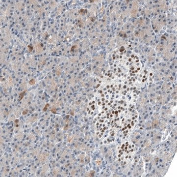
Immunohistochemistry Analysis: A 1:50 dilution from a representative lot detected MDM2 in human prostate cancer tissue.

Quality

Evaluated by Western Blot in A431 cell lysate.

Western Blot Analysis: 1 µg/mL of this antibody detected MDM2 in 10 µg of A431 cell lysate.

Target description

~85 kDa and ~60 kDa observed. Uniprot describes many isoforms between 12-56 kDa, which can be highly phosphorylated and ubiquitinated.

Physical form

Format: Purified
Protein G Purified
Purified mouse monoclonal IgG2bκ in buffer containing 0.1 M Tris-Glycine (pH 7.4), 150 mM NaCl with 0.05% sodium azide.

Storage and Stability

Stable for 1 year at 2-8°C from date of receipt.

Analysis Note

Control
A431 cell lysate

Other Notes

Concentration: Please refer to the Certificate of Analysis for the lot-specific concentration.

Disclaimer

Unless otherwise stated in our catalog or other company documentation accompanying the product(s), our products are intended for research use only and are not to be used for any other purpose, which includes but is not limited to, unauthorized commercial uses, in vitro diagnostic uses, ex vivo or in vivo therapeutic uses or any type of consumption or application to humans or animals.

Not finding the right product?  

Try our Product Selector Tool.

Storage Class Code

12 - Non Combustible Liquids

WGK

WGK 1

Flash Point(F)

Not applicable

Flash Point(C)

Not applicable


Certificates of Analysis (COA)

Search for Certificates of Analysis (COA) by entering the products Lot/Batch Number. Lot and Batch Numbers can be found on a product’s label following the words ‘Lot’ or ‘Batch’.

Already Own This Product?

Find documentation for the products that you have recently purchased in the Document Library.

Visit the Document Library

Eva Röijer et al.
The American journal of pathology, 160(2), 433-440 (2002-02-13)
Carcinoma ex pleomorphic adenoma (CexPA) is a carcinoma developing within a pre-existing benign pleomorphic adenoma (PA). Here we describe the identification and characterization of a series of genetic events leading to translocation, deletion/amplification, and overexpression of the HMGIC and MDM2
P Tanière et al.
British journal of cancer, 85(5), 721-726 (2001-09-05)
In Europe, high incidence rates of oesophageal squamous cell carcinoma (SCCE) are observed in western France (Normandy and Brittany) and in north-eastern Italy. Analysis of TP53 mutations in tumours from these regions has shown a high prevalence of mutations at
Robert F H Walter et al.
Journal of oncology, 2018, 1986982-1986982 (2018-08-17)
Previously, our group demonstrated that nuclear expression of E3 ubiquitin ligase (MDM2) in malignant pleural mesothelioma (MPM) is significantly associated with decreased overall survival. A possible explanation may be that overexpression of MDM2 leads to a proteasomal degradation of TP53
A Lakoma et al.
Cell death discovery, 1 (2015-01-01)
Neuroblastoma is an aggressive pediatric malignancy which is >98% p53 wild-type at diagnosis. As a primary repressor of p53 activity and part of a p53-activated negative feedback loop, targeting of mouse double minute 2 homolog (MDM2) is an attractive therapeutic
Marcin Poreba et al.
Cell death and differentiation, 26(12), 2695-2709 (2019-04-13)
Most caspases can be positioned unambiguously within the regulated cell death networks of apoptosis and pyroptosis, but the role of caspase-2, a highly conserved protease within the family, remains enigmatic. This is mainly due to lack of selective chemical and

Our team of scientists has experience in all areas of research including Life Science, Material Science, Chemical Synthesis, Chromatography, Analytical and many others.

Contact Technical Service