推荐产品
生物源
mouse
品質等級
100
500
共軛
unconjugated
抗體表格
culture supernatant
抗體產品種類
primary antibodies
無性繁殖
16/f5, monoclonal
描述
For In Vitro Diagnostic Use in Select Regions (See Chart)
形狀
buffered aqueous solution
物種活性
human
包裝
vial of 0.1 mL concentrate (376M-94)
vial of 0.5 mL concentrate (376M-95)
bottle of 1.0 mL predilute (376M-97)
vial of 1.0 mL concentrate (376M-96)
bottle of 7.0 mL predilute (376M-98)
製造商/商標名
Cell Marque™
技術
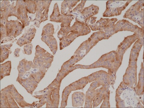
immunohistochemistry (formalin-fixed, paraffin-embedded sections): 1:100-1:500
同型
IgG1κ
控制
breast, pancreatic ductal adenocarcinoma, urothelial carcinoma
運輸包裝
wet ice
儲存溫度
2-8°C
視覺化
cytoplasmic, nuclear
基因資訊
human ... S100P(6286)
一般說明
S100P is a member of the S100 family of proteins. The family is expressed in a wide range of cells and is thought to play a role in cell cycle progression and in differentiation. S100P was initially identified in the placenta at rather high levels. Anti-S100P with nuclear or nuclear/ cytoplasmic immunoreactivity can be seen in essentially 100% of pancreatic ductal adenocarcinoma in pancreatic resection, and fine needle aspiration biopsy specimens. Anti-S100P displays no staining in the benign pancreatic ducts and acinar glands. S100P has been detected in the cells of virtually all intraductal papillary mucinous neoplasms tested. S100P is clearly expressed in the invasive component of intraductal papillary mucinous neoplasms (100%), including perineural, lymphatic, and minimal invasion. Biopsies of bile ducts with primary adenocarcinomas (90%) have exhibited strong nuclear and cytoplasmic staining for anti-S100P, with none of the 32 benign biopsies exhibiting anti-S100P immunoreactivity. An immunohistochemical panel including anti-S100P can be helpful in distinguishing adenocarcinoma from reactive epithelial changes on challenging bile duct biopsies. The detection of S100P expression may help distinguish urothelial carcinomas from other genitourinary neoplasms that enter into the differential diagnosis.
品質
 IVD |  IVD |  IVD |  RUO |
聯結
S100P Positive Control Slides, Product No. 376S, are available for immunohistochemistry (formalin-fixed, paraffin-embedded sections).
外觀
Solution in Tris Buffer, pH 7.3-7.7, with 1% BSA and <0.1% Sodium Azide
準備報告
Download the IFU specific to your product lot and formatNote: This requires a keycode which can be found on your packaging or product label.
其他說明
For Technical Service please contact: 800-665-7284 or email: service@cellmarque.com
法律資訊
Cell Marque is a trademark of Merck KGaA, Darmstadt, Germany
Not finding the right product?
Try our 产品选型工具.
Tatjana Crnogorac-Jurcevic et al.
The Journal of pathology, 201(1), 63-74 (2003-09-02)
In order to expand our understanding of the molecular changes underlying the complex pathology of pancreatic malignancy, global gene expression profiling of pancreatic adenocarcinoma compared with normal pancreatic tissue was performed. Human cDNA arrays comprising 9932 elements were interrogated with
Fan Lin et al.
The American journal of surgical pathology, 32(1), 78-91 (2007-12-29)
Recently, we demonstrated von Hippel-Lindau gene product (pVHL) was expressed in normal pancreatic ducts but absent in pancreatic ductal adenocarcinoma (PDA). Previous studies have suggested the diagnostic value of S100P, S100A4, and S100A6 in PDA. In this study, we evaluated
John P T Higgins et al.
The American journal of surgical pathology, 31(5), 673-680 (2007-04-27)
The morphologic distinction between prostate and urothelial carcinoma can be difficult. To identify novel diagnostic markers that may aid in the differential diagnosis of prostate versus urothelial carcinoma, we analyzed expression patterns in prostate and bladder cancer tissues using complementary
Kohei Nakata et al.
Human pathology, 41(6), 824-831 (2010-02-16)
Intraductal papillary mucinous neoplasms of the pancreas are subclassified based on morphological features, and different immunohistochemical profiles have been identified in association with the subtypes. We previously reported that S100P was an early developmental marker of pancreatic carcinogenesis and that
Mary Levy et al.
Human pathology, 41(9), 1210-1219 (2010-04-13)
Histopathologic distinction between benign and malignant bile duct epithelial lesions on endoscopic biopsies can be extremely challenging because of limited material, crush artifact, and compounding inflammatory and/or reactive changes particularly after stent placement. In this study, a total of 72
我们的科学家团队拥有各种研究领域经验,包括生命科学、材料科学、化学合成、色谱、分析及许多其他领域.
联系技术服务部门


