
A4187
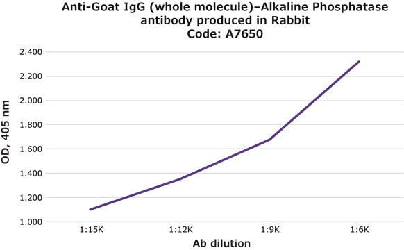
Anti-Goat IgG (whole molecule)–Alkaline Phosphatase antibody produced in rabbit
affinity isolated antibody, buffered aqueous glycerol solution
Synonym(s):
Rabbit Anti-Goat IgG (whole molecule)–AP
About This Item
Recommended Products


biological source
rabbit
conjugate
alkaline phosphatase conjugate
antibody form
affinity isolated antibody
antibody product type
secondary antibodies
clone
polyclonal
form
buffered aqueous glycerol solution
species reactivity
goat
technique(s)
direct ELISA: 1:30,000
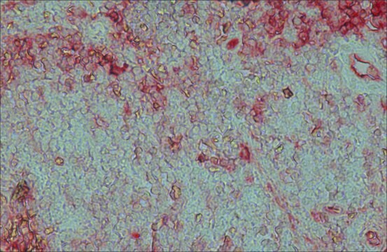
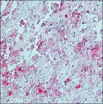
immunohistochemistry (formalin-fixed, paraffin-embedded sections): 1:50
western blot: 1:30,000
shipped in
wet ice
storage temp.
2-8°C
target post-translational modification
unmodified
Looking for similar products? Visit Product Comparison Guide
Related Categories
General description
Specificity
Immunogen
Application
Physical form
Disclaimer
Not finding the right product?
Try our Product Selector Tool.
Storage Class Code
10 - Combustible liquids
WGK
WGK 2
Choose from one of the most recent versions:
Certificates of Analysis (COA)
Don't see the Right Version?
If you require a particular version, you can look up a specific certificate by the Lot or Batch number.
Already Own This Product?
Find documentation for the products that you have recently purchased in the Document Library.
Customers Also Viewed


Our team of scientists has experience in all areas of research including Life Science, Material Science, Chemical Synthesis, Chromatography, Analytical and many others.
Contact Technical Service