Tropomyosin alpha-3 chain (UniProt: Q63610; also known as Gamma-tropomyosin, Tropomyosin-3, Tropomyosin-5) is encoded by the Tpm3 (also known as Tpm-5, Tpm5) gene (Gene ID: 117557) in rat. The TPM3 gene codes for the slow-twitch skeletal muscle isoform ( s Tm) and at least 9 LMW cytoskeletal isoforms referred to as Tm5NM1 to Tm5NM11. Tropomyosins are dimers of coiled-coil proteins that provide stability to actin filaments and regulate access of other actin-binding proteins. In muscle cells, they regulate muscle contraction by controlling the binding of myosin heads to the actin filament. In non-muscle cells tropomyosins are implicated in stabilizing cytoskeleton actin filaments. Tropomyosins have been implicated in the pathogenesis of cancer where high molecular weight isoforms are consistently down-regulated in transformed cells, while malignant cells display an increased reliance on low molecular weight isoforms. Tropomyosin 3 is a homodimeric protein that can form heterodimers with a beta (TPM2) chain. Its coiled coil structure is formed by 2 polypeptide chains. (Ref.: Glass, JJ et al. (2015). BMC Cancer. 15; 712).
Specificity
Clone 2G10.2 is a mouse monoclonal antibody that specifically detects Tpm3.1 and Tpm3.2. It targets an epitope within 27 amino acids from the C-terminal region.
Immunogen
Diphtheria Toxoid Carrier Protein-conjugated linear peptide corresponding to 27 amino acids from the C-terminal region of rat Tpm3.1 and Tpm3.2
Application
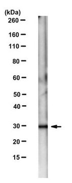
Western Blotting Analysis: 1 µg/mL from a representative lot detected Tropomyosin 3 in human lung tissue lysate.
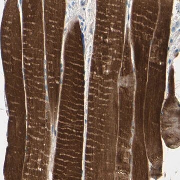
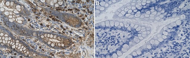
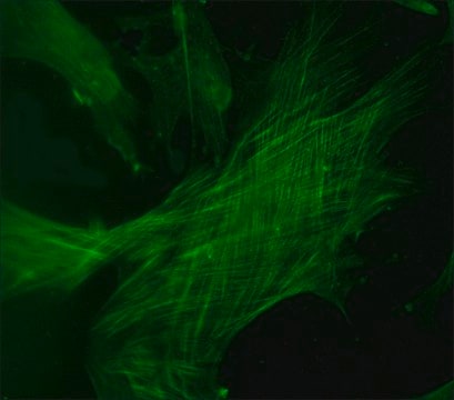
Immunofluorescence Analysis: A representative lot detected Tropomyosin 3 in Immunofluorescence applications (Schevzov, G., et. al. (2011). Bioarchitecture. 1(4):135-164).
Western Blotting Analysis: A representative lot detected Tropomyosin 3 in Western Blotting applications (Schevzov, G., et. al. (2011). Bioarchitecture. 1(4):135-164).
Quality
Evaluated by Western Blotting in mouse liver tissue lysate. Western Blotting Analysis: 1 µg/mL of this antibody detected Tropomyosin 3 in mouse liver tissue lysate.
Physical form
Purified mouse monoclonal antibody IgG2b in buffer containing 0.1 M Tris-Glycine (pH 7.4), 150 mM NaCl with 0.05% sodium azide.
Storage and Stability
Stable for 1 year at 2-8°C from date of receipt.
Other Notes
Concentration: Please refer to lot specific datasheet.
Disclaimer
Unless otherwise stated in our catalog or other company documentation accompanying the product(s), our products are intended for research use only and are not to be used for any other purpose, which includes but is not limited to, unauthorized commercial uses, in vitro diagnostic uses, ex vivo or in vivo therapeutic uses or any type of consumption or application to humans or animals.
Not finding the right product?
Try our Product Selector Tool.
Certificates of Analysis (COA)
Search for Certificates of Analysis (COA) by entering the products Lot/Batch Number. Lot and Batch Numbers can be found on a product’s label following the words ‘Lot’ or ‘Batch’.
Already Own This Product?
Find documentation for the products that you have recently purchased in the Document Library.
Our team of scientists has experience in all areas of research including Life Science, Material Science, Chemical Synthesis, Chromatography, Analytical and many others.