Skip to Content
Merck
All Photos(1)

Documents

MAB5234

Sigma-Aldrich

Anti-Neurocan Antibody

Chemicon®, from mouse

Sign Into View Organizational & Contract Pricing


About This Item

UNSPSC Code:
12352203
eCl@ss:
32160702
NACRES:
NA.41

biological source

mouse

Quality Level

antibody form

purified immunoglobulin

antibody product type

primary antibodies

clone

monoclonal

species reactivity

chicken

manufacturer/tradename

Chemicon®

technique(s)

ELISA: suitable
immunoprecipitation (IP): suitable
western blot: suitable

isotype

IgG1

NCBI accession no.

UniProt accession no.

shipped in

dry ice

target post-translational modification

unmodified

Gene Information

human ... NCAN(1463)

General description

Neurocan is the major soluble chondroitin sulfate proteoglycan in the brain. It is thought to play a functional role in axonal growth and guidance and in the establishment of specific neural pathways during embryonic brain development. Neurocan expression in the brain is developmentally regulated. Early on the major form of neurocan consists of a 245 kD core protein with approximately two chondroitin sulfate glycosoaminoglycan chains of 22 kD each. Later neurocan comprises a 180 kD core protein. Both forms of neurocan contain only chondroitin 4-sulfate glycosoaminoglycan chains. By virtue of their high expression at sites of neurnal damage and trauma, chondroitin sulfate proteoglycans, including neurocan, are thought to inhibit successful nerve regeneration.

Specificity

Neurocan, chicken. Recognizes the middle region of neurocan.

Application

Anti-Neurocan Antibody is an antibody against Neurocan for use in ELISA, IP & WB.
Western blot: <0.5 μg/mL

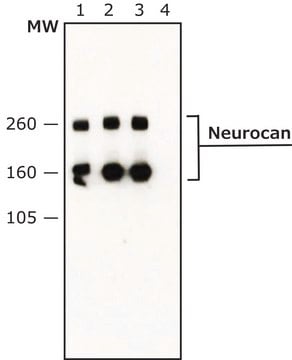
Western blot using anti-chick Neurocan (MAB5234). Samples are 1) Untreated embryonic chick brain extract, 2) chondroitinase-treated embryonic chick brain extract, 3) GST fusion proteins from the middle region of chick neurocan.

Samples must be digested with chondroitinase prior to running on SDS gels because undigested phosphacan is too large for most gels. Treatment is at a concentration of chondroitinase of 10U/mL in Tris-HCL pH 8.0. Make tissue or cell extract in 20-50mM Tris pH 7.6-8.0 with 0.15M NaCl in the presence of protease inhibitors. Add 1 microliter of enzyme to 30 microliters of extract and incubate 30 minutes at 37C. Then add SDS sample buffer, heat or boil sample as normal for SDS reducing samples.

Immunoprecipitation: 1 μg/mL

ELISA: 1 μg/mL, excellent for core protein, good for monomer

Immunocytochemistry: not tested

Immunohistochemistry: does not work on fixed samples, unfixed has not been tested.

Optimal working dilutions must be determined by the end user.

Physical form

Format: Purified

Other Notes

Concentration: Please refer to the Certificate of Analysis for the lot-specific concentration.

Legal Information

CHEMICON is a registered trademark of Merck KGaA, Darmstadt, Germany

Not finding the right product?  

Try our Product Selector Tool.

Storage Class Code

12 - Non Combustible Liquids

WGK

WGK 2

Flash Point(F)

Not applicable

Flash Point(C)

Not applicable


Certificates of Analysis (COA)

Search for Certificates of Analysis (COA) by entering the products Lot/Batch Number. Lot and Batch Numbers can be found on a product’s label following the words ‘Lot’ or ‘Batch’.

Already Own This Product?

Find documentation for the products that you have recently purchased in the Document Library.

Visit the Document Library

Polysialylated NCAM and ephrinA/EphA regulate synaptic development of GABAergic interneurons in prefrontal cortex.
Brennaman, LH; Zhang, X; Guan, H; Triplett, JW; Brown, A; Demyanenko, GP; Manis et al.
Cerebral Cortex (1991)
Keisuke Ikegami et al.
Scientific reports, 9(1), 13634-13634 (2019-09-22)
ST8 alpha-N-acetyl-neuraminide alpha-2,8-sialyltransferase 2 (ST8SIA2) synthesizes polysialic acid (PSA), which is essential for brain development. Although previous studies reported that St8sia2-deficient mice that have a mixed 129 and C57BL/6 (B6) genetic background showed mild and variable phenotypes, the reasons for
Rowena C Cua et al.
Glia, 61(6), 972-984 (2013-04-05)
Acute trauma to the central nervous system (CNS) can result in permanent damage and loss of function related to the poor regeneration of injured axons. Injured axons encounter several barriers to regeneration, such as the glial scar at the injury
Shane A Liddelow et al.
Nature, 541(7638), 481-487 (2017-01-19)
Reactive astrocytes are strongly induced by central nervous system (CNS) injury and disease, but their role is poorly understood. Here we show that a subtype of reactive astrocytes, which we termed A1, is induced by classically activated neuroinflammatory microglia. We

Our team of scientists has experience in all areas of research including Life Science, Material Science, Chemical Synthesis, Chromatography, Analytical and many others.

Contact Technical Service