General description
Proliferating cell nuclear antigen (UniProt: P04961; also known as PCNA) is encoded by the Pcna gene (Gene ID: 25737) in rat. PCNA is a homotrimeric nuclear protein that serves as an auxiliary protein of DNA polymerase delta and is involved in the control of eukaryotic DNA replication by increasing the polymerase′s processability during elongation of the leading strand. It Induces a robust stimulatory effect on the 3′-5′ exonuclease and 3′-phosphodiesterase, but not apurinic-apyrimidinic (AP) endonuclease, APEX2 activities. It is reported to colocalize with CREBBP, EP300 and POLD1 to sites of DNA damage. It acts as a loading platform to recruit DDR proteins that allow completion of DNA replication after DNA damage and promote post-replication repair. PCNA can undergo phosphorylation on tyrosine 211 by EGFR that stabilizes chromatin-associated PCNA. Following DNA damage, it can undergo monoubiquitination to stimulate direct bypass of DNA lesions by specialized DNA polymerases or polyubiquitination to promote recombination-dependent DNA synthesis across DNA lesions by template switching mechanisms. Mutations in PCN gene are known to cause Ataxia-telangiectasia-like disorder 2 (ATLD2) that is characterized by developmental delay, ataxia, hearing loss, and short stature.
Specificity
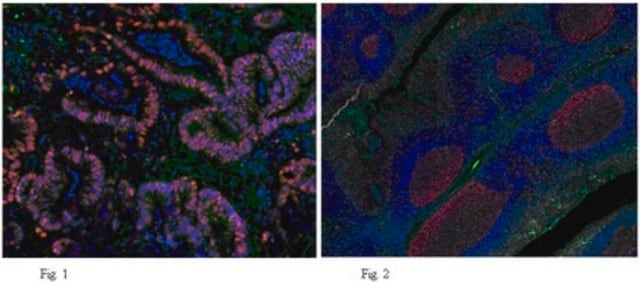
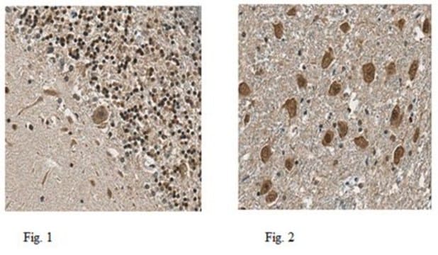
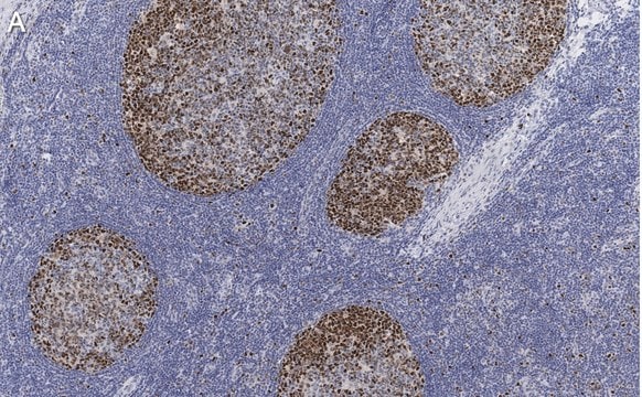
Clone PC10 recognizes PCNA from all vertebrate and insect species tested and has been used to identify transformed cells (Kurki, 1988), proliferating cells in solid tumors (Smetana, 1983), and blast cells in leukemia patients (Takasaki, 1984). Immunohistochemistry with clone PC10 has been used to study the expression of PCNA in paraffin sections of normal tissues and lymphoid neoplasms (Hall, 1990). In proliferating cells, PCNA staining pattern is predominantly nuclear. By western blot, the antibody detects a polypeptide migrating at 36 kDa with an isoelectric point of 4.8.
Immunogen
Rat PCNA induced in the Protein A expression vector pR1T2T.
Application
Anti-PCNA Antibody, clone PC10 is a mouse monoclonal antibody for detection of PCNA also known as Proliferating Cell Nuclear Antigen, DNA Polymerase delta Processivity Factor & has been validated in ELISA, FC, IP, WB,IHC(P).
Research Category
Epigenetics & Nuclear Function
Research Sub Category
Cell Cycle, DNA Replication & Repair
Physical form
Format: Purified
The monoclonal is presented at a concentration of 100μg/1mL in phosphate buffered saline containing 10mM sodium azide and 1mg/ml bovine serum albumin. We recommend that each laboratory determine an optimum working titre for use in its particular application.
Storage and Stability
For use within 1 month of purchase store at +4°C, for long term storage aliquot antibody into small volumes and store at -20°C. Avoid repeated freeze/ thawing.
Analysis Note
Control
POSITIVE CONTROL: Tonsil or reactive lymph node.
Other Notes
Concentration: Please refer to the Certificate of Analysis for the lot-specific concentration.
Legal Information
CHEMICON is a registered trademark of Merck KGaA, Darmstadt, Germany
Disclaimer
Unless otherwise stated in our catalog or other company documentation accompanying the product(s), our products are intended for research use only and are not to be used for any other purpose, which includes but is not limited to, unauthorized commercial uses, in vitro diagnostic uses, ex vivo or in vivo therapeutic uses or any type of consumption or application to humans or animals.
