
M6068
Anti-phospho-Myosin Light Chain (pSer19) antibody produced in rabbit
affinity isolated antibody, buffered aqueous glycerol solution
Synonym(s):
Anti-MLC
Sign Into View Organizational & Contract Pricing
All Photos(1)
About This Item
Pricing and availability is not currently available.
Recommended Products


biological source
rabbit
Quality Level
conjugate
unconjugated
antibody form
affinity isolated antibody
antibody product type
primary antibodies
clone
polyclonal
form
buffered aqueous glycerol solution
species reactivity
human (predicted), chicken
technique(s)
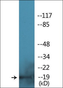
western blot: 1:1,000 using chicken gizzard myosin II phosphorylated in vitro by PAK
shipped in
dry ice
storage temp.
−20°C
target post-translational modification
phosphorylation (pSer19)
Gene Information
human ... MYL1(4632)
Related Categories
General description
Myosin light chains belongs to the calmodulin (CaM) gene family.
Rabbit polyclonal anti-phospho-Myosin Light Chain (pSer19) antibody detects chicken MLC. Results show that reactivity with the antibody requires phosphorylation of MLC by active kinase, and that only the peptide corresponding to MLC (pSer19) blocks the antibody signal, thereby demonstrating the specificity of the antibody. Human (100% homologous) has not been tested, but is expected to react.
Immunogen
synthetic phosphopeptide derived from the region of chicken MLC that contains serine 19 (serine 20 including the initiating methionine). Human (100% homologous) has not been tested, but is expected to cross-react.
Application
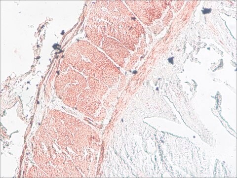
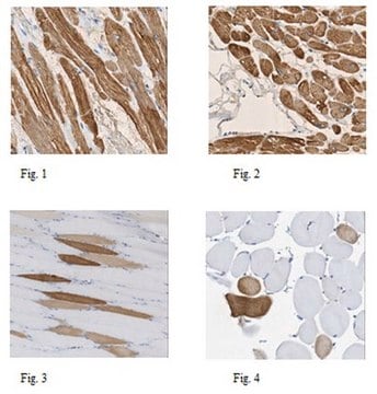
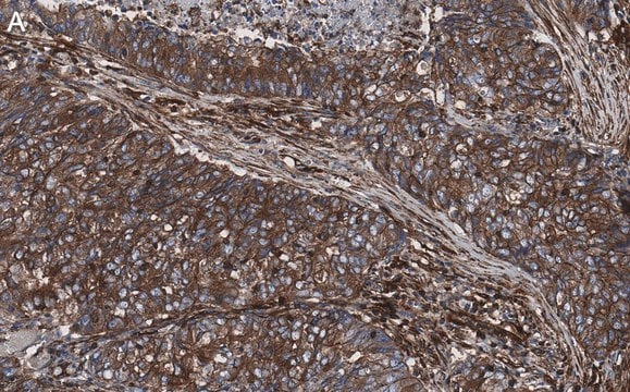
Anti-phospho-Myosin Light Chain (pSer19) antibody produced in rabbit has been used in the detection of phosphorylated myosin: in human lung fibroblasts and avian lens cells by immunoblotting by immunohistochemical detection in rabbit aorta samplesby immunofluorescence detection in human submandibular gland tissues
Biochem/physiol Actions
Myosin light chain is a subunit of the conventional myosins, e.g., myosin II. In smooth muscle and nonmuscle cells, conventional myosins mediate a wide variety of contractile events including cytokinesis, cell motility, and smooth muscle contraction. MLC is phosphorylated by multiple serine-threonine kinases such as Rho-kinase and PAK, however, myosin light chain kinase (MLCK) acts as the primary kinase. Contractile activity of conventional myosins is regulated by phosphorylation of MLC on several residues. Phosphorylation of serine 19, the major phosphorylation site and the preferred site for MLCK, activates myosin motor activity under physiological conditions.
Myosin light chains functions to maintain integrity of myosin protein. Mutations in the myosin light chain is implicated in familial hypertrophic cardiomyopathy.
Physical form
Solution in Dulbecco′s phosphate buffered saline (without Mg2+ and Ca2+), pH 7.3 (± 0.1), 50% glycerol with 1.0 mg/mL BSA (IgG and protease free) and 0.05% sodium azide.
Disclaimer
Unless otherwise stated in our catalog or other company documentation accompanying the product(s), our products are intended for research use only and are not to be used for any other purpose, which includes but is not limited to, unauthorized commercial uses, in vitro diagnostic uses, ex vivo or in vivo therapeutic uses or any type of consumption or application to humans or animals.
Not finding the right product?
Try our Product Selector Tool.
Storage Class Code
10 - Combustible liquids
WGK
WGK 1
Flash Point(F)
Not applicable
Flash Point(C)
Not applicable
Choose from one of the most recent versions:
Already Own This Product?
Find documentation for the products that you have recently purchased in the Document Library.
The effects of actomyosin disruptors on the mechanical integrity of the avian crystalline lens
Won GJ et al.
Molecular Vision, 21(9), 98-98 (2015)
Shuichi Asano et al.
Physiological reports, 5(9) (2017-05-17)
In patients with pulmonary diseases such as idiopathic pulmonary fibrosis and severe acute respiratory distress syndrome, progressive pulmonary fibrosis is caused by dysregulated wound healing via activation of fibroblasts after lung inflammation or severe damage. Migration of fibroblasts toward the
Matrix stiffness regulates migration of human lung fibroblasts
Asano S, et al.
Physiological Reports, 5(9), e13281-e13281 (2017)
Dorian Obino et al.
Cell reports, 25(11), 3110-3122 (2018-12-13)
Complete activation of B cells relies on their capacity to extract tethered antigens from immune synapses by either exerting mechanical forces or promoting their proteolytic degradation through lysosome secretion. Whether antigen extraction can also be tuned by local cues originating
Myosin essential light chain in health and disease
Hernandez OM, et al.
American Journal of Physiology. Heart and Circulatory Physiology, 292(4), H1643-H1654 (2007)
Active Filters
Our team of scientists has experience in all areas of research including Life Science, Material Science, Chemical Synthesis, Chromatography, Analytical and many others.
Contact Technical Service