Skip to Content
Merck
All Photos(1)

Key Documents

SRP3221

Sigma-Aldrich

M-CSF from mouse

Animal-component free, recombinant, expressed in E. coli, ≥98% (SDS-PAGE), ≥98% (HPLC), suitable for cell culture

Synonym(s):

CSF-1, MGI-IM, Macrophage Colony Stimulating Factor

Sign Into View Organizational & Contract Pricing


About This Item

UNSPSC Code:
12352202
NACRES:
NA.32

biological source

mouse

recombinant

expressed in E. coli

Assay

≥98% (HPLC)
≥98% (SDS-PAGE)

form

lyophilized

potency

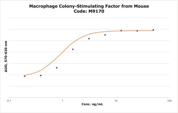
≤10 ng/mL ED50

mol wt

36.4 kDa

packaging

pkg of 10 μg

technique(s)

cell culture | mammalian: suitable

impurities

<0.1 EU/μg endotoxin, tested

color

white to off-white

UniProt accession no.

shipped in

wet ice

storage temp.

−20°C

Gene Information

mouse ... CSF1(12977)

General description

M-CSF (macrophage colony-stimulating factor) is a potent hematopoietic factor produced by a variety of cells including lymphocytes, monocytes, fibroblasts, endothelial cells, myoblasts and osteoblasts. The protein works through its receptor CSF1R (colony stimulating factor 1 receptor). The human is reactive in murine systems, but the murine molecule exhibits no activity on human cells. Recombinant murine M-CSF is a 36.4kDa homodimeric protein containing two 156 amino acid polypeptide subunits.

Biochem/physiol Actions

M-CSF (macrophage colony-stimulating factor) is a key regulator of cellular proliferation, differentiation, and survival of blood monocytes, tissue macrophages and their progenitor cells. M-CSF has been shown to play important roles in modulating dermal thickness, and male and female fertility. It is clinically used in the treatment of infection, malignancies and atherosclerosis. It facilitates hematopoietic recovery after bone marrow transplantation. M-CSF also works as a pleiotropic growth factor and plays a crucial role in reproduction and organogenesis. Absence of it can result in reduced growth, neurological and reproductive problems as well as developmental defects in the mammary gland, bone and pancreas.
M-CSF is a potent hematopoietic factor produced by a variety of cells including lymphocytes, monocytes, fibroblasts, endothelial cells, myoblasts and osteoblasts. Recombinant murine M-CSF is a 36.4 kDa homodimeric protein containing two 156 amino acid polypeptide subunits.

Sequence

MKEVSEHCSH MIGNGHLKVL QQLIDSQMET SCQIAFEFVD QEQLDDPVCY LKKAFFLVQD IIDETMRFKD NTPNANATER LQELSNNLNS CFTKDYEEQN KACVRTFHET PLQLLEKIKN FFNETKNLLE KDWNIFTKNC NNSFAKCSSR DVVTKP

Physical form

10 mM sodium phosphate, 50 mM sodium chloride, pH 7.5.

Reconstitution

Centrifuge the vial prior to opening. Reconstitute in water to a concentration of 0.1-0.5 mg/ml. Do not vortex. This solution can be stored at 2-8°C for up to 1 week. For extended storage, it is recommended to further dilute in a buffer containing a stabilizer (example 5% Trehalose) and store in working aliquots at -20°C to -80°C.

Pictograms

Exclamation mark

Signal Word

Warning

Hazard Statements

Hazard Classifications

Eye Irrit. 2 - Skin Irrit. 2 - STOT SE 3

Target Organs

Respiratory system

Storage Class Code

13 - Non Combustible Solids

WGK

WGK 3

Flash Point(F)

Not applicable

Flash Point(C)

Not applicable


Certificates of Analysis (COA)

Search for Certificates of Analysis (COA) by entering the products Lot/Batch Number. Lot and Batch Numbers can be found on a product’s label following the words ‘Lot’ or ‘Batch’.

Already Own This Product?

Find documentation for the products that you have recently purchased in the Document Library.

Visit the Document Library

GuiHao Chen et al.
Journal of molecular and cellular cardiology, 159, 62-79 (2021-06-18)
Monocytes and macrophages are cellular forces that drive and resolve inflammation triggered by acute myocardial ischemia. One of the most important but least understood regulatory mechanisms is how these cells sense cues from the micro-milieu and integrate environmental signals with
OSMR deficiency aggravates pressure overload-induced cardiac hypertrophy by modulating macrophages and OSM/LIFR/STAT3 signalling.
Feng, et al.
Journal of Translational Medicine, 21, 290-290 (2023)
Chenming Wu et al.
Immunology and cell biology, 97(1), 72-84 (2018-09-13)
The role of specific histone deacetylase (HDAC) proteins in regulating the lipopolysaccharide (LPS)-induced inflammatory response and its underlying mechanisms are unclear. Here, HDAC2, a class I HDAC family protein, is essential for the LPS-triggered inflammatory response in macrophages. LPS stimulation
Targeted disruption of the mouse colony-stimulating factor 1 receptor gene results in osteopetrosis, mononuclear phagocyte deficiency, increased primitive progenitor cell frequencies, and reproductive defects.
Dai XM, et al.
Blood, 99, 111-120 (2002)
Jin Liu et al.
Nature communications, 13(1), 7519-7519 (2022-12-07)
Regulatory T cells (Tregs) are critically involved in neovascularization, an important compensatory mechanism in peripheral artery disease. The contribution of G protein coupled receptor 174 (GPR174), which is a regulator of Treg function and development, in neovascularization remains elusive. Here

Our team of scientists has experience in all areas of research including Life Science, Material Science, Chemical Synthesis, Chromatography, Analytical and many others.

Contact Technical Service