Skip to Content
Merck
All Photos(2)

Documents

ABC432

Sigma-Aldrich

Anti-LC3B Antibody

serum, from rabbit

Synonym(s):

Microtubule-associated proteins 1A/1B light chain 3B, Autophagy-related protein LC3 B, Autophagy-related ubiquitin-like modifier LC3 B, MAP1 light chain 3-like protein 2, MAP1A/MAP1B light chain 3 B, MAP1A/MAP1B LC3 B, Microtubule-associated protein 1 li

Sign Into View Organizational & Contract Pricing


About This Item

UNSPSC Code:
12352203
eCl@ss:
32160702
NACRES:
NA.41

biological source

rabbit

Quality Level

antibody form

serum

antibody product type

primary antibodies

clone

polyclonal

species reactivity

human

technique(s)

western blot: suitable

NCBI accession no.

UniProt accession no.

shipped in

wet ice

target post-translational modification

unmodified

Gene Information

General description

MAP1LC3B, also known as Autophagy-related protein LC3 B, LC3B, Autophagy-related ubiquitin-like modifier LC3 B, MAP1 light chain 3-like protein 2, MAP1A/MAP1B light chain 3 B, MAP1A/MAP1B LC3 B, Microtubule-associated protein 1 light chain 3 beta, and encoded by the gene MAP1LC3B/MAP1ALC3, is a ubiquitin-like modifier involved in formation of autophagosomal vacuoles (autophagosomes). MAP1LC3B plays a role in mitophagy which contributes to regulate mitochondrial quantity and quality by eliminating the mitochondria to a basal level to fulfill cellular energy requirements and preventing excess ROS production. Whereas LC3s are involved in elongation of the phagophore membrane, the GABARAP/GATE-16 subfamily is essential for a later stage in autophagosome maturation. MAP1LC3B interacts at microtubules with CABP1 (via EF-hands 1 and 2) but not with calmodulin. MAP1LC3B interacts with FYCO1 (via C-terminus). MAP1LC3B interacts with TP53INP1 and TP53INP2, and MAP1LC3B interacts with TBC1D25. MAP1LC3B directly interacts with SQSTM1; this interaction leads to MAP1LC3B recruitment to inclusion bodies containing polyubiquitinated protein aggregates and to inclusion body degradation by autophagy. MAP1LC3B interacts with ATG4B, MAPK15 and BNIP3 protein as well. MAP1LC3B is localized to the cytoplasm and cytoskeleton membranes, specifically endomembranes and cytoplasmic vesicles in the autophagosome. MAP1LC3B is expressed in most abundantly in heart, brain, skeletal muscle and testis tissues, with very low expression in liver tissues. Interestingly, high expression of MAP1LC3B in triple negative breast cancer (TNBC) is associated with poor outcomes in TNBC patients and is currently being studied as a prognostic evaluation marker for patients with TNBC.

Immunogen

Linear peptide corresponding to human LC3B.

Application

Detect MAP1 using this rabbit polyclonal antibody, Anti-LC3B Antibody validated for use in western blotting.
Research Category
Apoptosis & Cancer
Research Sub Category
Apoptosis - Additional
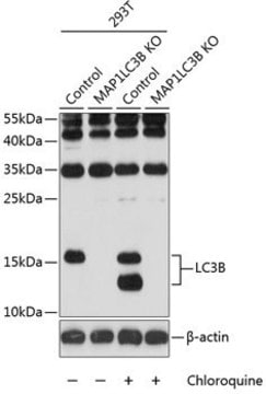
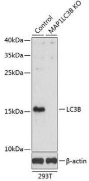
Western Blotting Analysis: A representative lot from an independent laboratory detected LC3B in shRNA control HeLa cell lysates and demonstrated a loss of signal in TBC1D5 knockdown HeLa cell lysates. HeLa cells were either serum starved, or treated with bafilomycin A1 or serum starved in the presence of bafilomycin A1 (Popovic, D., et al. (2012). Mol Cell Biol. 32(9):1733-1744.).
Immunohistochemistry (Paraffin) Analysis: A 1:500 dilution from a representative lot detected LC3B in human cerebral cortex and rat cerebral cortex tissue sections.

Quality

Evaluated by Western Blotting in HeLa cell lysate.

Western Blotting Analysis: A 1:2,000 dilution of this antibody detected LC3B in 10 µg of HeLa cell lysate.

Target description

~15 kDa observed

Physical form

Rabbit polyclonal serum with 0.05% sodium azide.
Unpurified

Storage and Stability

Stable for 1 year at -20°C from date of receipt.
Handling Recommendations: Upon receipt and prior to removing the cap, centrifuge the vial and gently mix the solution. Aliquot into microcentrifuge tubes and store at -20°C. Avoid repeated freeze/thaw cycles, which may damage IgG and affect product performance.

Disclaimer

Unless otherwise stated in our catalog or other company documentation accompanying the product(s), our products are intended for research use only and are not to be used for any other purpose, which includes but is not limited to, unauthorized commercial uses, in vitro diagnostic uses, ex vivo or in vivo therapeutic uses or any type of consumption or application to humans or animals.

Not finding the right product?  

Try our Product Selector Tool.

Storage Class Code

10 - Combustible liquids

WGK

WGK 1


Certificates of Analysis (COA)

Search for Certificates of Analysis (COA) by entering the products Lot/Batch Number. Lot and Batch Numbers can be found on a product’s label following the words ‘Lot’ or ‘Batch’.

Already Own This Product?

Find documentation for the products that you have recently purchased in the Document Library.

Visit the Document Library

Ping Qi et al.
Oncology letters, 12(6), 4577-4583 (2017-01-20)
It has been well-established that apoptosis contributes to cancer cell death; however, the role of autophagy in cancer cell death remains unclear. The aim of the present study was to investigate the effects of rottlerin, a traditional Indian medicine, on
Ying Zhang et al.
Cell death & disease, 13(12), 1034-1034 (2022-12-13)
Although second-generation therapies like abiraterone (ABI) and enzalutamide (ENZ) benefit patients with castration-resistant prostate cancer (CRPC), drug resistance frequently occurs, eventually resulting in therapy failure. In this study, we used two libraries, FDA-approved drug library and CRISP/Cas9 knockout (GeCKO) library
Yuanyuan Yang et al.
Cell death & disease, 11(1), 39-39 (2020-01-22)
Ginsenosides exhibit a large variety of biological activities in maintaining physical health; however, the molecule underpinnings underlining these biological activities remain to be defined. Here, we took a cellular condition that compound K (CK) induces autophagic cell death in HeLa
Ai-Gang Sun et al.
Molecular medicine reports, 16(6), 7939-7948 (2017-10-07)
CDGSH iron sulfur domain 2 (CISD2) has been found to be important in carcinogenesis. However, the role of CISD2 in glioma remains to be elucidated. The present study aimed to investigate the role of CISD2 in glioma using the reverse

Our team of scientists has experience in all areas of research including Life Science, Material Science, Chemical Synthesis, Chromatography, Analytical and many others.

Contact Technical Service