Skip to Content
Merck
All Photos(3)

Key Documents

AB5622-I

Sigma-Aldrich

Anti-MAP-2

from rabbit, purified by affinity chromatography

Synonym(s):

Mirotubule associated protein 2

Sign Into View Organizational & Contract Pricing


About This Item

UNSPSC Code:
12352203
eCl@ss:
32160702
NACRES:
NA.41

biological source

rabbit

Quality Level

antibody form

affinity isolated antibody

antibody product type

primary antibodies

clone

polyclonal

purified by

affinity chromatography

species reactivity

rat, mouse

technique(s)

immunocytochemistry: suitable
immunohistochemistry: suitable (paraffin)
western blot: suitable

NCBI accession no.

UniProt accession no.

shipped in

ambient

target post-translational modification

unmodified

Gene Information

rat ... Map2(25595)

General description

Microtubule-associated protein 2 (UniProt: P15146; also known as MAP-2) is encoded by the Map2 (also known as Mtap2) gene (Gene ID: 25595) in Rat. MAPs are a family of proteins that bind to and stabilize microtubules. MAP4 is expressed ubiquitously while MAP1 and MAP2 are expressed primarily in neurons. MAP2 are cytoskeletal proteins that stabilize microtubules and prevents their depolymerization and may also have a stiffening effect on microtubules. MAP-2 plays an important role in brain neuron microtubule assembly. It also associates with neurofilaments and actin filaments and thereby guides interaction among microtubules, other cytoskeletal elements, and cytoplasmic organelles. Four different isoforms of MAP2 have been described that bind to microtubules through a domain near the carboxyl terminus. MAP2 isoforms are expressed only in neuronal cells and specifically in the cell bodies and dendrites. In the central nervous system, MAP-2 is confined to neuronal cell bodies and dendrites. However, some axons may also have small amounts of MAP-2. MAP-2 usually consists of MAP-2a and MAP-2b, ~280kDa each, and lower molecular weight MAP2c, which is present during neuronal differentiation in the developing nervous system. MAP-2 is a marker for neurons and is uniformly distributed throughout the cell when first expressed in cultured neurons. However, later it is selectively localized as dendritic development proceeds. MAP-2 can be phosphorylated at serine residues in K-X-G-S motifs by MAP/microtubule affinity-regulating kinase (MARK1 or MARK2) that results in detachment from microtubules, and their disassembly.

Specificity

This polyclonal antibody detects MAP2 in Mouse and Rat tissues.

Immunogen

KLH-conjugated three peptides from N-and C-terminal regions of rat MAP-2.

Application

Anti-MAP-2, Cat. No. AB5622-I, is a highly specific rabbit polyclonal antibody that targets microtubule-associated protein 2 and has been tested in Immunocytochemistry, Immunohistochemistry (Paraffin), and Western Blotting.
Immunohistochemistry Analysis: A 1:1,000 dilution from a representative lot detected MAP-2 in rat cerebral cortex and rat kidney tissue.

Western Blotting Analysis: 2 µg/mL from a representative lot detected MAP-2 in 10 µg of rat brain microsomal preparations.

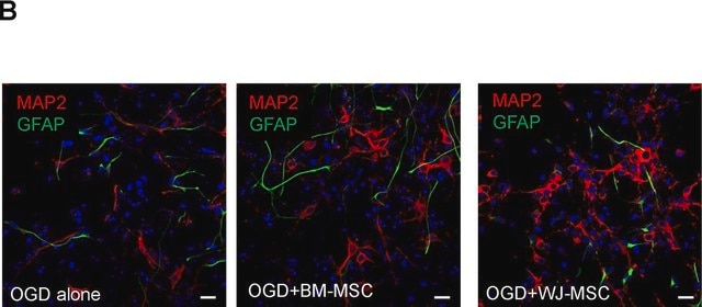
Immunocytochemistry Analysis: 1 µg/mL from a representative lot detected MAP-2 in rat primary cortex cells.
Research Category
Neuroscience

Quality

Evaluated by Immunohistochemistry in mouse cerebellum tissue.

Immunohistochemistry Analysis: A 1:1,000 dilution of this antibody detected MAP-2 in mouse cerebellum tissue.

Target description

~280 kDa observed; 202.41 kDa calculated. Uncharacterized bands may be observed in some lysate(s).

Physical form

Affinity Purified
Purified rabbit polyclonal antibody in buffer containing 0.1 M Tris-Glycine (pH 7.4), 150 mM NaCl with 0.05% sodium azide.

Storage and Stability

Stable for 1 year at 2-8°C from date of receipt.

Other Notes

Concentration: Please refer to lot specific datasheet.

Disclaimer

Unless otherwise stated in our catalog or other company documentation accompanying the product(s), our products are intended for research use only and are not to be used for any other purpose, which includes but is not limited to, unauthorized commercial uses, in vitro diagnostic uses, ex vivo or in vivo therapeutic uses or any type of consumption or application to humans or animals.

Not finding the right product?  

Try our Product Selector Tool.

Storage Class Code

12 - Non Combustible Liquids

WGK

WGK 1

Flash Point(F)

Not applicable

Flash Point(C)

Not applicable


Certificates of Analysis (COA)

Search for Certificates of Analysis (COA) by entering the products Lot/Batch Number. Lot and Batch Numbers can be found on a product’s label following the words ‘Lot’ or ‘Batch’.

Already Own This Product?

Find documentation for the products that you have recently purchased in the Document Library.

Visit the Document Library

Anna Karpova et al.
Cellular and molecular life sciences : CMLS, 80(8), 228-228 (2023-07-26)
Nuclear Ca2+ waves elicited by NMDAR and L-type voltage-gated Ca2+-channels as well as protein transport from synapse-to-nucleus are both instrumental in control of plasticity-related gene expression. At present it is not known whether fast [Ca2+]n transients converge in the nucleus
Avik Roy et al.
Journal of Alzheimer's disease : JAD, 84(4), 1747-1762 (2021-11-09)
Doublecortin (DCX), a microtubule associated protein, has emerged as a central biomarker of hippocampal neurogenesis. However, molecular mechanisms by which DCX is regulated are poorly understood. Since sleep is involved with the acquisition of memory and oleamide or 9-Octadecenamide (OCT)
Noriko Koganezawa et al.
The European journal of neuroscience, 53(11), 3548-3560 (2021-04-15)
Effective drugs that can cure cognitive impairments remain elusive. Because synaptic dysfunction has been correlated with cognitive impairments, drug development to target synaptic dysfunction is important. Recently, natural compounds and crude drugs have emerged as potential therapeutic agents for cognitive
Nicholas C Vierra et al.
eLife, 8 (2019-10-31)
The voltage-gated K+ channel Kv2.1 serves a major structural role in the soma and proximal dendrites of mammalian brain neurons, tethering the plasma membrane (PM) to endoplasmic reticulum (ER). Although Kv2.1 clustering at neuronal ER-PM junctions (EPJs) is tightly regulated
Mohammed S Basabrain et al.
Bioengineering (Basel, Switzerland), 9(11) (2022-11-11)
Cell-based neural regeneration is challenging due to the difficulty in obtaining sufficient neural stem cells with clinical applicability. Stem cells from apical papilla (SCAPs) originating from embryonic neural crests with high neurogenic potential could be a promising cell source for

Our team of scientists has experience in all areas of research including Life Science, Material Science, Chemical Synthesis, Chromatography, Analytical and many others.

Contact Technical Service