Protein PD-1, mPD-1, CD279) is encoded by the PDCD1 (also known as PD1) gene (Gene ID: 18566) in murine species. PD-1 is a monomeric inhibitory cell surface receptor involved in the regulation of T-cell function during immunity and tolerance. PD-1 is synthesized with a signal peptide (aa 1-20), which is subsequently cleaved off. The mature form contains an extracellular domain (aa 21-169), a transmembrane domain (aa 170-190), and a cytoplasmic domain (aa 191-288). PD-1 also contains a single N-terminal immunoglobulin variable region (IgV) like domain (aa 31-139). PD-1 and its ligands, PD-L1 and PD-L2 play a key role in the maintenance of peripheral tolerance, a process by which the quiescence of autoreactive mature T cells is maintained. However, tumors and pathogens that cause chronic infections can exploit this pathway to escape T-cell mediated tumor-specific and pathogen-specific immunity. The effector functions of T-cells expressing PD-1 can be downregulated by PD-L1 or PD-L2 expressed by the tumor cells. PD-1 lacks SH2- or SH3-binding motifs on its cytoplasmic tail, but contains the N-terminal sequence VDYGEL that forms an immunoreceptor tyrosine-based inhibition motif (V/I/LxYxxL), which recruits SH2 domain-containing phosphatases. The cytoplasmic tail also contains the C-terminal sequence TEYATI, which forms an immunoreceptor tyrosine-based switch motif (TxYxxL). PD-1 ligation is reported to inhibit the activation of T-cell receptor proximal kinases, which results in attenuation of Lck-mediated phosphorylation of ZAP-70 and initiation of downstream events. PD-1 is also reported to impair the activation of the MEK-ERK MAP kinase pathway by inhibiting activation of PLC- 1 and Ras.
Specificity
Clone G4 is an Armenian Hamster monoclonal antibody that specifically detects PD-1 in murine cells.
Immunogen
Murine PD-1Ig fusion protein.
Application
Affects Function Analysis: A representative lot detected PD-1 in Affects Funtion applications (Dronca, R.S., et. al. (2016). JCI Insight. 1(6); Hirano, F., et. al. (2005). Cancer Res. 65(3):1089-96; Overacre-Delgoffe, A.E., et. al. (2017). Cell. 169(6):1130; Tsushima, F., et. al. (2006). Blood. 110(1):180-5).
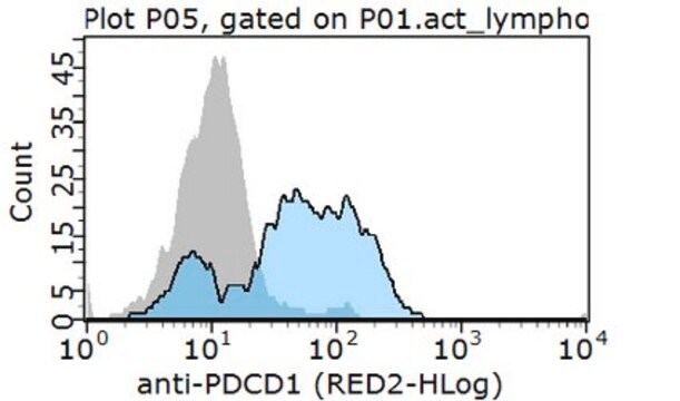
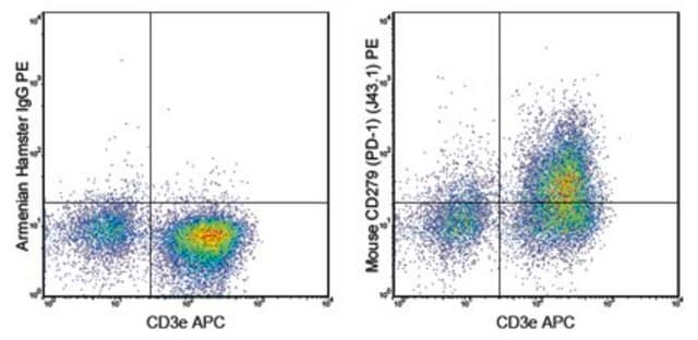
Flow Cytometry Analysis: A representative lot detected PD-1 in Flow Cytometry applications (Hirano, F., et. al. (2005). Cancer Res. 65(3):1089-96).
Detects Programmed cell death protein 1 using this armenian hamster monoclonal Anti-PD-1, clone G4, Cat. No. MABC1132, testted for use in Flow Cytometry and Affects Function.
Quality
Evaluated by Flow Cytometry in EL4 T lymphoma cells.
Flow Cytometry Analysis: 1 µg of this antibody detected PD-1 in 1X10E6 EL4 T lymphoma cells.
Target description
~31.84 kDa calculated.
Physical form
Format: Purified
Other Notes
Concentration: Please refer to lot specific datasheet.
Not finding the right product?
Try our Product Selector Tool.
Certificates of Analysis (COA)
Search for Certificates of Analysis (COA) by entering the products Lot/Batch Number. Lot and Batch Numbers can be found on a product’s label following the words ‘Lot’ or ‘Batch’.
Already Own This Product?
Find documentation for the products that you have recently purchased in the Document Library.
Cancer immunology research, 10(2), 162-181 (2021-12-17)
Cytotoxic CD8+ T cells (CTL) are a crucial component of the immune system notable for their ability to eliminate rapidly proliferating malignant cells. However, the T-cell intrinsic factors required for human CTLs to accomplish highly efficient antitumor cytotoxicity are not
Although the immune checkpoint role of programmed death ligand 1 (PD-L1) has been established and targeted in cancer immunotherapy, the tumor-intrinsic role of PD-L1 is less appreciated in tumor biology and therapeutics development, partly because of the incomplete mechanistic understanding.
Questions
Reviews
★★★★★ No rating value
Active Filters
Our team of scientists has experience in all areas of research including Life Science, Material Science, Chemical Synthesis, Chromatography, Analytical and many others.