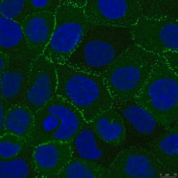
This antibody shows distinct punctate membrane staining of desmoplakins 1 (MW 250 kDa) and 2 (MW 215 kDa) in stratified epithelia and simple epithelia, including glands, urothelium, thymic reticular epithelium, hepatocytes, intercalated disks of myocardium and arachnoid cells of meninges.
Immunogen
Bovine desmoplakin 1 & 2
Application
Anti-Desmoplakin 1& 2 Antibody, clone DP2.15 detects level of Desmoplakin 1& 2 & has been published & validated for use in IF, WB & IC.
Research Category Cell Structure
Research Sub Category Cytoskeleton
Western blot Immunohistochemistry: 1:10; Frozen tissue Immunofluorescence
Optimal working dilutions must be determined by the end user.
Physical form
Format: Purified
Lyophilized. Reconstitute in 1 mL distilled water (final solution contains 0.09% sodium azide, 0.5% BSA in PBS buffer, pH 7.4
Storage and Stability
Reconstituted antibody can be stored at 2°-8°C for up to 12 months from date of receipt. Antibody can also be stored at -20°C in undiluted aliquots.
Legal Information
CHEMICON is a registered trademark of Merck KGaA, Darmstadt, Germany
Disclaimer
Unless otherwise stated in our catalog or other company documentation accompanying the product(s), our products are intended for research use only and are not to be used for any other purpose, which includes but is not limited to, unauthorized commercial uses, in vitro diagnostic uses, ex vivo or in vivo therapeutic uses or any type of consumption or application to humans or animals.
Not finding the right product?
Try our Product Selector Tool.
Storage Class Code
12 - Non Combustible Liquids
WGK
WGK 2
Flash Point(F)
Not applicable
Flash Point(C)
Not applicable
Certificates of Analysis (COA)
Search for Certificates of Analysis (COA) by entering the products Lot/Batch Number. Lot and Batch Numbers can be found on a product’s label following the words ‘Lot’ or ‘Batch’.
Already Own This Product?
Find documentation for the products that you have recently purchased in the Document Library.
The physiological functions of microtubules (MTs) are poorly understood in many differentiated cell types. We developed a genetic toolkit to study MT dynamics and function in diverse cells. Using TRE-EB1-GFP mice, we found that MT dynamics are strongly suppressed in
Cellular and molecular gastroenterology and hepatology, 13(4), 1181-1200 (2021-12-21)
Desmosomes are intercellular junctions connecting keratin intermediate filaments of neighboring cells. The cadherins desmoglein 2 (Dsg2) and desmocollin 2 mediate cell-cell adhesion, whereas desmoplakin (Dsp) provides the attachment of desmosomes to keratins. Although the importance of the desmosome-keratin network is
Desmosomes are cell-cell junctions that provide mechanical integrity to epithelial and cardiac tissues. Desmosomes have two distinct adhesive states, calcium-dependent and hyperadhesive, which balance tissue plasticity and strength. A highly ordered array of cadherins in the adhesive interface is hypothesized
Basal stem cells fuel development, homeostasis, and regeneration of the epidermis. The proliferation and fate decisions of these cells are highly regulated by their microenvironment, including the basement membrane and underlying mesenchymal cells. Basal progenitors give rise to differentiated progeny
International journal of molecular sciences, 22(21) (2021-11-14)
Aging is the major risk factor for cardiovascular disease, which is the leading cause of mortality worldwide among aging populations. Cisd2 is a prolongevity gene that mediates lifespan in mammals. Previously, our investigations revealed that a persistently high level of
Questions
Reviews
★★★★★ No rating value
Active Filters
Our team of scientists has experience in all areas of research including Life Science, Material Science, Chemical Synthesis, Chromatography, Analytical and many others.