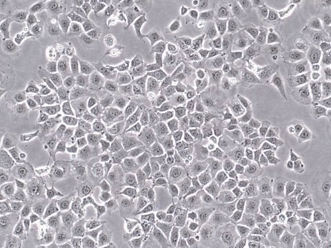
SCC177
TS/A Mouse Mammary Adenocarcinoma Cell Line
Mouse
Synonym(s):
TSA
Sign Into View Organizational & Contract Pricing
All Photos(2)
About This Item
UNSPSC Code:
41106514
eCl@ss:
32011203
NACRES:
NA.81
Recommended Products
product name
TS/A Mouse Mammary Adenocarcinoma Cell Line, TS/A mouse mammary adenocarcinoma cell line is a well characterized and highly published model for breast cancer research.
biological source
mouse
technique(s)
cell culture | mammalian: suitable
shipped in
ambient
General description
Metastasizing (stage IV) breast cancers have the lowest survival rate among all stages of breast cancer. Metastases may take on a range of morphologies in their new locations, complicating diagnosis and decisions on courses of treatment. Tumor cell lines that mimic the in vivo behavior of metastasizing cancers are thus critical tools in breast cancer research.
TS/A is a highly metastatic mouse mammary adenocarcinoma cell line exhibiting a marked degree of morphological heterogeneity in culture, with a range of differentiated cell types from epithelial-like to fibroblast-like (1). TS/A cells express estrogen receptor and colony-stimulating factor and are negative for the adhesion protein ICAM-1 (2,3). The TS/A cell line is a well-characterized and highly published model for tumor metastasis and heterogeneity and is a popular tool for studies of immunological gene therapy (4,5). It may also be used as a syngeneic mouse model of breast cancer.
The TS/A cell line was isolated from a mammary adenocarcinoma that arose spontaneously in a BALB/c female mouse (1).
The TS/A cell line was isolated from a mammary adenocarcinoma that arose spontaneously in a BALB/c female mouse (1).
Cell Line Description
Cancer Cells
Application
Research Category
Cancer
Oncology
Cancer
Oncology
This product is intended for sale and sold solely to academic institutions for internal academic research use per the terms of the “Academic Use Agreement” as detailed in the product documentation. For information regarding any other use, please contact licensing@emdmillipore.com.
Quality
• Each vial contains ≥ 1X10⁶ viable cells.
• Cells are tested negative for infectious diseases by a Mouse Essential CLEAR panel by Charles River Animal Diagnostic Services.
• Cells are verified to be of mouse origin and negative for inter-species contamination from rat, chinese hamster, Golden Syrian hamster, human and non-human primate (NHP) as assessed by a Contamination CLEAR panel by Charles River Animal Diagnostic Services.
• Cells are negative for mycoplasma contamination.
Disclaimer
Unless otherwise stated in our catalog or other company documentation accompanying the product(s), our products are intended for research use only and are not to be used for any other purpose, which includes but is not limited to, unauthorized commercial uses, in vitro diagnostic uses, ex vivo or in vivo therapeutic uses or any type of consumption or application to humans or animals.
Storage Class Code
12 - Non Combustible Liquids
WGK
WGK 2
Flash Point(F)
does not flash
Flash Point(C)
does not flash
Certificates of Analysis (COA)
Search for Certificates of Analysis (COA) by entering the products Lot/Batch Number. Lot and Batch Numbers can be found on a product’s label following the words ‘Lot’ or ‘Batch’.
Already Own This Product?
Find documentation for the products that you have recently purchased in the Document Library.
A Allione et al.
Cancer research, 54(23), 6022-6026 (1994-12-01)
To evaluate the efficacy of vaccinations with cytokine-gene-transduced tumor cells, BALB/c mice were challenged with 1 x 10(5) parental cells of a syngeneic adenocarcinoma cell line (TSA-pc). No protection was observed in mice immunized 30 days earlier with 1 x
G Nicoletti et al.
British journal of cancer, 52(2), 215-222 (1985-08-01)
We investigated the presence of colony-stimulating factor (CSF) in supernatants obtained from TS/A, a new metastatic murine cell line, and from its high-and low-metastatic clonal derivatives (E and F clones, respectively). TS/A cells produced a CSF in vitro that induced
P Nanni et al.
Clinical & experimental metastasis, 1(4), 373-380 (1983-10-01)
A metastasizing mouse cell line (TS/A), originated from a mammary adenocarcinoma which arose spontaneously in a BALB/c female retired breeder, has been established in vitro. It displayed a remarkable morphologic heterogeneity, which is evident in plastic adherent cultures (cell types
P Musiani et al.
Laboratory investigation; a journal of technical methods and pathology, 74(1), 146-157 (1996-01-01)
Impressive inhibition of tumor growth has been observed after transduction of cytokine genes into tumor cells. Secreted cytokines do not affect the proliferation of a tumor directly but activate a host immune reaction strong enough to overcome its oncogenic capacity.
Ai Sato et al.
STAR protocols, 2(2), 100488-100488 (2021-05-28)
Here, we describe an immunofluorescence (IF) microscopy-based approach to quantify cytosolic double-stranded DNA molecules in cultured eukaryotic cells upon the selective and specific permeabilization of plasma membranes. This technique is compatible with widefield microscopy coupled with automated image analysis for
Our team of scientists has experience in all areas of research including Life Science, Material Science, Chemical Synthesis, Chromatography, Analytical and many others.
Contact Technical Service


