Skip to Content
Merck
All Photos(3)

Documents

AB10523

Sigma-Aldrich

Anti-EBF-1 Antibody

from rabbit, purified by affinity chromatography

Synonym(s):

Transcription factor COE1, O/E-1, OE-1, Early B-cell factor

Sign Into View Organizational & Contract Pricing


About This Item

UNSPSC Code:
12352203
eCl@ss:
32160702
NACRES:
NA.41

biological source

rabbit

Quality Level

antibody form

affinity isolated antibody

antibody product type

primary antibodies

clone

polyclonal

purified by

affinity chromatography

species reactivity

human, mouse

technique(s)

dot blot: suitable
immunofluorescence: suitable
immunohistochemistry: suitable (paraffin)
western blot: suitable

NCBI accession no.

UniProt accession no.

shipped in

wet ice

target post-translational modification

unmodified

Gene Information

human ... EBF1(1879)

General description

The family of Early B-cell Factor (EBF) or Collier/Olf-1/EBF (COE) proteins, EBF1, EBF2, and EBF3, are ubiquitous zinc-binding transcription factors found in many species, including human, Drosophila melanogaster, and Caenorhabditis elegans. EBF1 is abundantly expressed in early B-cells, lymph node, spleen, and adipose tissue. Low levels of EBF1 have also been detected in the brain, heart, skeletal muscle, and kidney. EBF proteins are capable of forming homo- and heterodimers that bind to DNA at specific sites that include the sequence 5′-ATTCCCNNGGGAATT-3′. EBF1, the prototypical member of this family, may employ epigenetic mechanisms, such as DNA methylation and chromatin remodeling, to regulate genes involved in B-cell development such as Pax5. The second member of this family EBF2 is involved in bone development, adipogenesis, and CNS development, processes which may also hinge on EBF1. EBF3 has been characterized as a tumor suppressor protein, due to its role in regulating multiple genes such as those for cyclins and CDKs which control cell growth, differentiation, and apoptosis. However, emerging evidence indicates that all EBF proteins may play a role in the development of various cancers.

Specificity

This antibody recognizes EBF-1 but not EBF-2 or EBF-3.

Immunogen

KLH-conjugated linear peptide corresponding to mouse EBF-1.

Application

Anti-EBF-1 Antibody is an antibody against EBF-1 for use in Western Blotting, IHC(P), Immunofluorescence, Dot Blot.
Research Category
Neuroscience
Research Sub Category
Developmental Neuroscience
Western Blot Analysis: 2 µg/mL from a representative lot detected EBF-1 in COS-7 cells transfected with EBF-3. (Image courtesy of Dr. Giacomo Consalez, San Raffaele Scientific Institute.)

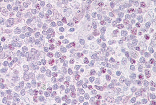
Immunohistochemistry Analysis: A 1:500 dilution from a representative lot detected EBF-1 in mouse cryosections of wild type spinal cord tissue. (Image courtesy of Dr. Giacomo Consalez, San Raffaele Scientific Institute.)

Immunofluorescence Analysis: A 1:500 dilution from a representative lot detected EBF-1 in COS-7 cells transfected with EBF expressing plasmids. (Image courtesy of Dr. Giacomo Consalez, San Raffaele Scientific Institute.)

Dot Blot Analysis: EBF-1, EBF-2, and EBF-3 peptides from a representative lot were probed with Anti-EBF-1 (1:100 dilution). No cross reactivity to peptides for EBF-3 & EBF-2 were observed.

Quality

Evaluated by Western Blot in Raji cell lysate.

Western Blot Analysis: 2 µg/mL of this antibody detected EBF-1 in 10 µg of Raji cell lysate.

Target description

~66 kDa observed. Uncharacterized bands may appear between ~40 and ~170 kDa in some lysates.

Physical form

Affinity purified
Purified rabbit polyclonal in buffer containing 0.1 M Tris-Glycine (pH 7.4), 150 mM NaCl with 0.05% sodium azide.

Storage and Stability

Stable for 1 year at 2-8°C from date of receipt.

Analysis Note

Control
Raji cell lysate

Other Notes

Concentration: Please refer to the Certificate of Analysis for the lot-specific concentration.

Disclaimer

Unless otherwise stated in our catalog or other company documentation accompanying the product(s), our products are intended for research use only and are not to be used for any other purpose, which includes but is not limited to, unauthorized commercial uses, in vitro diagnostic uses, ex vivo or in vivo therapeutic uses or any type of consumption or application to humans or animals.

Not finding the right product?  

Try our Product Selector Tool.

Storage Class Code

12 - Non Combustible Liquids

WGK

WGK 1

Flash Point(F)

Not applicable

Flash Point(C)

Not applicable


Certificates of Analysis (COA)

Search for Certificates of Analysis (COA) by entering the products Lot/Batch Number. Lot and Batch Numbers can be found on a product’s label following the words ‘Lot’ or ‘Batch’.

Already Own This Product?

Find documentation for the products that you have recently purchased in the Document Library.

Visit the Document Library

Xiaolei Song et al.
Neuroscience bulletin, 37(7), 985-998 (2021-05-21)
Medium spiny neurons (MSNs) in the striatum, which can be divided into D1 and D2 MSNs, originate from the lateral ganglionic eminence (LGE). Previously, we reported that Six3 is a downstream target of Sp8/Sp9 in the transcriptional regulatory cascade of
Zhenmeiyu Li et al.
Cell death discovery, 8(1), 301-301 (2022-07-01)
The striatum is the main input structure of the basal ganglia, receiving information from the cortex and the thalamus and consisting of D1- and D2- medium spiny neurons (MSNs). D1-MSNs and D2-MSNs are essential for motor control and cognitive behaviors
Andrea Tinterri et al.
The Journal of comparative neurology, 526(3), 397-411 (2017-09-19)
In mammals, thalamic axons are guided internally toward their neocortical target by corridor (Co) neurons that act as axonal guideposts. The existence of Co-like neurons in non-mammalian species, in which thalamic axons do not grow internally, raised the possibility that
Agnieszka Szymula et al.
PLoS pathogens, 14(2), e1006890-e1006890 (2018-02-21)
The Epstein-Barr virus (EBV) nuclear antigen leader protein (EBNA-LP) is the first viral latency-associated protein produced after EBV infection of resting B cells. Its role in B cell transformation is poorly defined, but it has been reported to enhance gene
Paloma Merchan-Sala et al.
The Journal of comparative neurology, 525(13), 2805-2819 (2017-05-05)
The striatum is the major component of the basal ganglia and is well known to play a key role in the control of motor function via balanced output from the indirect (iSPNs) and direct pathway striatal projection neurons (dSPNs). Little

Our team of scientists has experience in all areas of research including Life Science, Material Science, Chemical Synthesis, Chromatography, Analytical and many others.

Contact Technical Service