Skip to Content
Merck
All Photos(3)

Key Documents

T3287

Sigma-Aldrich

Monoclonal Anti-Talin antibody produced in mouse

clone 8d4, ascites fluid

Synonym(s):

Monoclonal Anti-Talin

Sign Into View Organizational & Contract Pricing


About This Item

MDL number:
UNSPSC Code:
12352203
NACRES:
NA.41

biological source

mouse

Quality Level

conjugate

unconjugated

antibody form

ascites fluid

antibody product type

primary antibodies

clone

8d4, monoclonal

mol wt

antigen 225-235 kDa

contains

15 mM sodium azide

species reactivity

chicken, frog, rat, human, mouse

technique(s)

immunoprecipitation (IP): suitable
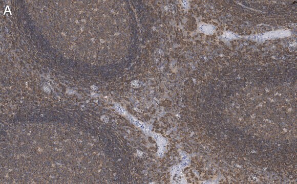
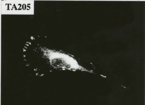
indirect immunofluorescence: 1:500 using cultured Rat2 cells
western blot: 1:100 using chicken gizzard extract

isotype

IgG1

UniProt accession no.

shipped in

dry ice

storage temp.

−20°C

target post-translational modification

unmodified

Gene Information

Looking for similar products? Visit Product Comparison Guide

General description

Monoclonal Anti-Talin (mouse IgG1 isotype) is derived from the 8d4 hybridoma produced by the fusion of mouse myeloma cells and splenocytes from immunized BALB/c mice. Talin is localized at a subset of adherens junctions, specialized cell-cell and cell-matrix associations that are characterized by the presence of filamentous actin at the cytoplasmic face of the junctional complex. In cultured cells, talin is absent from cell-cell junctions and found predominantly at adhesion plaques and in fibrillar streaks underlying cell surface fibronectin.
Talin 1 (TLN1) gene mapped to human chromosome 9p13.3, codes for a 270kDa cytoskeletal protein containing 2541 amino acids. Talin-1 is widely expressed in the kidney, liver, spleen, stomach, lung and vascular smooth muscle. The TLN1 antiparallel dimeric structure is characterized with a globular head containing four-point-one, ezrin, radixin, moesin (FERM) domain and rod domain involved in dimer formation.

Specificity

Reacts specifically with mammalian and avian talin. In immunoblotting, the antibody recognizes an epitope located on the 225 kDa chicken talin molecule and the 190 kDa fragment obtained by protease cleavage. It also localizes the slightly higher molecular weight talin in human platelets. The antibody labels focal adhesions, membrane ruffles, and ventral streaks in methanol/acetone fixed cultured fibroblasts.

Immunogen

purified chicken gizzard talin.

Application

Monoclonal Anti-Talin may be used for the localization of talin using various immunochemical assays such as immunoblot, immunocytochemistry, immunofluorescence, enzyme linked immunosorbent assay (ELISA) and for microinjection and transfection studies. The antibody recognizes native talin and can be used for immunoprecipitation.

Biochem/physiol Actions

Talin 1 (TLN1) is a cytoskeletal protein, which plays a vital role in cancer cell adhesion, migration and invasion. It is also involved in integrin activation and it facilitates the adhesion of integrin to actin cytoskeleton. TLN1 might act as a potential prognostic biomarker in nasopharyngeal carcinoma (NPC). Overexpression of TLN1 is observed in endometrioid carcinoma, oral squamous cell carcinoma (OSCC), prostate cancer and hepatocellular carcinoma. Serum TLN1 functions as a potential biomarker of hepatocellular carcinoma (HCC). Talin 1 (TLN1) is highly expressed in glioblastoma multiforme (GBM). The Inhibition of TLN1 activity helps in diminishing malignant features of GBM cells. It also plays an important role in regulation of, Bevacizumab (anti-angiogenic agent)-resistance.
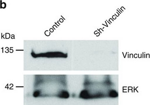
Talin interacts with at least two other proteins that are localized at adhesion plaques, vinculin and integrin. Talin and vinculin have been shown to interact with each other and both have been proposed to be involved in generating the transmembrane connection, between the extracellular matrix and the cytoskeleton, that occurs at adhesion plaques.

Disclaimer

Unless otherwise stated in our catalog or other company documentation accompanying the product(s), our products are intended for research use only and are not to be used for any other purpose, which includes but is not limited to, unauthorized commercial uses, in vitro diagnostic uses, ex vivo or in vivo therapeutic uses or any type of consumption or application to humans or animals.

Not finding the right product?  

Try our Product Selector Tool.

Storage Class Code

12 - Non Combustible Liquids

WGK

WGK 2

Flash Point(F)

Not applicable

Flash Point(C)

Not applicable


Certificates of Analysis (COA)

Search for Certificates of Analysis (COA) by entering the products Lot/Batch Number. Lot and Batch Numbers can be found on a product’s label following the words ‘Lot’ or ‘Batch’.

Already Own This Product?

Find documentation for the products that you have recently purchased in the Document Library.

Visit the Document Library

Regionally-selective cell colonization of micropatterned surfaces prepared by plasma polymerization of acrylic acid and 1, 7-octadiene.
Filova E, et al.
Physiological Research, 58(5) (2009)
CBAP functions as a novel component in chemokine-induced ZAP70-mediated T-cell adhesion and migration
Chiang YJ, et al.
PLoS ONE, 8(4), e61761-e61761 (2013)
Vassiliki Kostourou et al.
Nature communications, 4, 2020-2020 (2013-06-27)
Genetic ablation of endothelial focal adhesion kinase (FAK) can inhibit pathological angiogenesis, suggesting that loss of endothelial FAK is sufficient to reduce neovascularization. Here we show that reduced stromal FAK expression in FAK-heterozygous mice unexpectedly enhances both B16F0 and CMT19T
Ayman Alsebaey et al.
Asian Pacific journal of cancer prevention : APJCP, 17(8), 4077-4082 (2016-09-21)
Hepatocellular carcinoma (HCC) is a dreadful complication of end stage liver disease with high morbidity and mortality. The aim was to assess the role of serum talin-1 and non-invasive brosis in patients with HCC. A total of eighty seven subjects
Ming-Tsung Lai et al.
The Journal of pathology, 224(3), 367-376 (2011-05-07)
Oral squamous cell carcinoma (OSCC) is highly invasive and is associated with frequent tumour recurrences and lymph node metastases. Identification of genes involved in the aggressiveness of OSCC may provide new targets for clinical intervention. A genome-wide study based on

Our team of scientists has experience in all areas of research including Life Science, Material Science, Chemical Synthesis, Chromatography, Analytical and many others.

Contact Technical Service