Skip to Content
Merck
All Photos(2)

Key Documents

SAB4200462

Sigma-Aldrich

Anti-Claudin-1 antibody produced in rabbit

~1.0 mg/mL, affinity isolated antibody

Synonym(s):

Anti-CLD1, Anti-CLDN1, Anti-ILVASC, Anti-SEMP1

Sign Into View Organizational & Contract Pricing


About This Item

UNSPSC Code:
12352203
NACRES:
NA.41
conjugate:
unconjugated
application:
IHC
WB
clone:
polyclonal
species reactivity:
human, rat
citations:
10
technique(s):
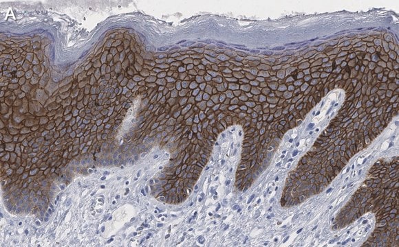
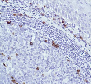
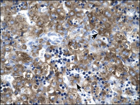
immunohistochemistry: 20 μg/mL using formalin-fixed, paraffin-embedded rat kidney.
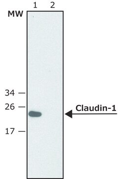
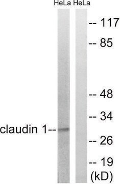
western blot: 1.5-3.0 μg/mL using extracts of rat kidney (S1 fraction).

biological source

rabbit

Quality Level

conjugate

unconjugated

antibody form

affinity isolated antibody

antibody product type

primary antibodies

clone

polyclonal

form

buffered aqueous solution

mol wt

antigen ~23 kDa

species reactivity

human, rat

concentration

~1.0 mg/mL

technique(s)

immunohistochemistry: 20 μg/mL using formalin-fixed, paraffin-embedded rat kidney.
western blot: 1.5-3.0 μg/mL using extracts of rat kidney (S1 fraction).

UniProt accession no.

shipped in

dry ice

storage temp.

−20°C

target post-translational modification

unmodified

Gene Information

human ... CLDN1(9076)
rat ... Cldn1(65129)

General description

Anti-Claudin-1 is produced in rabbit using as immunogen a synthetic peptide corresponding to a sequence located at the C-terminal region of human claudin-1, conjugated to KLH. Claudin-1 (also known as CLDN1, senescence-associated epithelial membrane protein (SEMP1) and ILVASC) is a 22 kDa protein. It is expressed at high levels in kidney and liver and at lower levels in spleen, heart, brain, lung and testis. Claudins are located in both epithelial and endothelial cells in tissues. Claudins consist of four transmembrane domains and two extracellular loops, required to form tight junction strands. CLDN1 gene is located on human chromosome 3q28.

Immunogen

synthetic peptide corresponding to a sequence located at the C-terminal region of human claudin-1, conjugated to KLH. The corresponding sequence is identical in rat claudin-1 and highly conserved (single amino acid substitution) in mouse claudin-1.

Application

Anti-Claudin-1 antibody produced in rabbit has been used in immunohistochemistry and immunoblotting,

Biochem/physiol Actions

Claudin-1 is frequently up-regulated in colorectal carcinomas (CRCs), resulting in tumor differentiation and progression. Defects in the gene encoding claudin-1 are the cause of an autosomal recessive syndrome named ichthyosis-sclerosing cholangitis neonatal (NISCH). It participates in the barrier formation of tight junction. It modulates the tight junctions in human airway epithelium. CLDN1 acts as a receptor for the hepatitis C virus entry.

Physical form

Solution in 0.01 M phos­phate buffered saline, pH 7.4, containing 15 mM sodium azide.

Disclaimer

Unless otherwise stated in our catalog or other company documentation accompanying the product(s), our products are intended for research use only and are not to be used for any other purpose, which includes but is not limited to, unauthorized commercial uses, in vitro diagnostic uses, ex vivo or in vivo therapeutic uses or any type of consumption or application to humans or animals.

Not finding the right product?  

Try our Product Selector Tool.

Storage Class Code

10 - Combustible liquids

Flash Point(F)

Not applicable

Flash Point(C)

Not applicable


Choose from one of the most recent versions:

Certificates of Analysis (COA)

Lot/Batch Number

Don't see the Right Version?

If you require a particular version, you can look up a specific certificate by the Lot or Batch number.

Already Own This Product?

Find documentation for the products that you have recently purchased in the Document Library.

Visit the Document Library

Claudin association with CD81 defines hepatitis C virus entry
Harris HJ, et al.
The Journal of biological chemistry, jbc-M110 (2010)
Taurine supplementation alleviates puromycin aminonucleoside damage by modulating endoplasmic reticulum stress and mitochondrial-related apoptosis in rat kidney
Stacchiotti A, et al.
Nutrients, 10(6), 689-689 (2018)
Tracking the fate of glomerular epithelial cells in vivo using serial multiphoton imaging in new mouse models with fluorescent lineage tags
Hackl MJ, et al.
Nature Medicine, 19(12), 1661-1661 (2013)
Contribution of tight junction proteins to ion, macromolecule, and water barrier in keratinocytes
Kirschner N, et al.
The Journal of Investigative Dermatology, 133(5), 1161-1169 (2013)
Claudin-1 regulates cellular transformation and metastatic behavior in colon cancer
Dhawan P, et al.
The Journal of Clinical Investigation, 115(7), 1765-1776 (2005)

Our team of scientists has experience in all areas of research including Life Science, Material Science, Chemical Synthesis, Chromatography, Analytical and many others.

Contact Technical Service