L9006
Lectin from Ulex europaeus (gorse, furze)
FITC conjugate, lyophilized powder
Synonym(s):
Ulex europaeus agglutinin, UEA-I
Sign Into View Organizational & Contract Pricing
All Photos(1)
About This Item
UNSPSC Code:
12352202
NACRES:
NA.32
Recommended Products
conjugate
FITC conjugate
Quality Level
form
lyophilized powder
potency
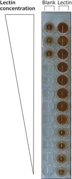
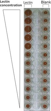
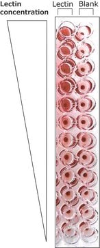
<5 μg/mL agglutination activity
composition
Protein, ~10% Lowry
extent of labeling
2-5 mol FITC per mol protein
storage temp.
−20°C
Looking for similar products? Visit Product Comparison Guide
Related Categories
Biochem/physiol Actions
Ulex europaeus agglutinin has anti-H blood group specificity. There are two types of lectin: UEA I has an affinity for L-fucose and UEA II has an affinity for N,N′-diacetylchitobiose. The mol. wt. of UEA I was initially found to be 170,000 daltons. Later reports indicate that UEA I may form aggregates at neutral and basic pH and that the correct mol. wt. is 68,000 daltons.
Packaging
Package size based on protein content.
Linkage
Conjugates are prepared from affinity purified lectin.
Physical form
Contains potassium phosphate buffer salts and NaCl
Analysis Note
Agglutination activity is expressed in μg/ml and is determined from serial dilutions in phosphate buffered saline, pH 6.8, of a 1 mg/ml solution. This activity is the lowest concentration to agglutinate a 2% suspension of human blood group O erythrocytes after 1 hour incubation at 25 °C.
Storage Class Code
11 - Combustible Solids
WGK
WGK 3
Flash Point(F)
Not applicable
Flash Point(C)
Not applicable
Personal Protective Equipment
dust mask type N95 (US), Eyeshields, Gloves
Choose from one of the most recent versions:
Certificates of Analysis (COA)
Lot/Batch Number
Don't see the Right Version?
If you require a particular version, you can look up a specific certificate by the Lot or Batch number.
Already Own This Product?
Find documentation for the products that you have recently purchased in the Document Library.
Customers Also Viewed
Karla Lehle et al.
Artificial organs, 40(6), 577-585 (2015-10-30)
Multipotent progenitor cells were mobilized during pediatric extracorporeal membrane oxygenation (ECMO). We hypothesize that these cells also adhered onto polymethylpentene (PMP) fibers within the membrane oxygenator (MO) during adult ECMO support. Mononuclear cells were removed from the surface of explanted
Naoki Akanuma et al.
Pancreas, 46(9), 1202-1207 (2017-09-14)
We aimed to evaluate the contribution of acinar-to-ductal metaplasia (ADM) to the accumulation of cells with a ductal phenotype in cultured human exocrine pancreatic tissues and reveal the underlying mechanism. We sorted and cultured viable cell populations in human exocrine
Alan Pestronk et al.
Muscle & nerve, 42(1), 53-61 (2010-06-15)
The causes of perifascicular myofiber atrophy and capillary pathology in dermatomyositis are incompletely understood. We studied 11 dermatomyositis muscles by histochemistry, immunohistochemistry, and ultrastructure. We found that endomysial capillaries within regions of perifascicular atrophy are not entirely lost, but they
Zoe E Clayton et al.
International journal of cardiology, 234, 81-89 (2017-02-18)
Endothelial cells derived from human induced pluripotent stem cells (iPSC-ECs) promote angiogenesis, and more recently induced endothelial cells (iECs) have been generated via fibroblast trans-differentiation. These cell types have potential as treatments for peripheral arterial disease (PAD). However, it is
Gang Chen et al.
The Journal of clinical investigation, 119(10), 2914-2924 (2009-09-18)
Various acute and chronic inflammatory stimuli increase the number and activity of pulmonary mucus-producing goblet cells, and goblet cell hyperplasia and excess mucus production are central to the pathogenesis of chronic pulmonary diseases. However, little is known about the transcriptional
Our team of scientists has experience in all areas of research including Life Science, Material Science, Chemical Synthesis, Chromatography, Analytical and many others.
Contact Technical Service


