Skip to Content
Merck
All Photos(1)

Key Documents

H2287

Sigma-Aldrich

Anti-Histone Deacetylase 6 (HDAC6) antibody produced in rabbit

IgG fraction of antiserum, buffered aqueous solution

Synonym(s):

Anti-CPBHM, Anti-HD6, Anti-JM21, Anti-PPP1R90

Sign Into View Organizational & Contract Pricing


About This Item

MDL number:
UNSPSC Code:
12352203
NACRES:
NA.41
conjugate:
unconjugated
application:
ARR
IP
WB
clone:
polyclonal
species reactivity:
human, mouse
citations:
12
technique(s):
immunoprecipitation (IP): 5-10 μg using RIPA extract of 500 μg of 293T cells expressing recombinant mouse HDAC6.
microarray: suitable
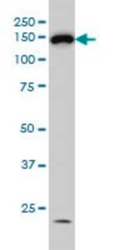
western blot: 1:1,000 using 293T cells expressing recombinant mouse HDAC6 in a chemiluminescent detection system.

biological source

rabbit

Quality Level

conjugate

unconjugated

antibody form

IgG fraction of antiserum

antibody product type

primary antibodies

clone

polyclonal

form

buffered aqueous solution

mol wt

antigen ~134 kDa

species reactivity

human, mouse

technique(s)

immunoprecipitation (IP): 5-10 μg using RIPA extract of 500 μg of 293T cells expressing recombinant mouse HDAC6.
microarray: suitable
western blot: 1:1,000 using 293T cells expressing recombinant mouse HDAC6 in a chemiluminescent detection system.

UniProt accession no.

shipped in

dry ice

storage temp.

−20°C

target post-translational modification

unmodified

Gene Information

human ... HDAC6(10013)
mouse ... Hdac6(15185)

General description

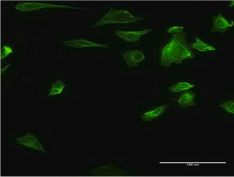
Anti-Histone Deacetylase 6 (HDAC6), is produced in rabbit using a synthetic peptide corresponding to amino acid residues 1199-1213 of human HDAC6. Mammalian HDACs can be divided into three classes according to sequence homology. Class I consists of the yeast Rpd3-like proteins HDAC1, HDAC2, HDAC3 and HDAC8. Class II consists of the yeast Hda1-like proteins HDAC4, HDAC5, HDAC6, HDAC7, HDAC9 and HDAC10. Class III consists of the yeast Sir2-like proteins. Class I HDACs are ubiquitously expressed, most class II HDACs are tissue-specific. Class II HDACs are larger than those of class I and their catalytic domain is located in the carboxy-terminal half of the protein. HDAC6 is a unique deacetylase among class II members in that it contains a duplicate of the catalytic domain in its NH2 terminus as well. The deacetylase activity of class II HDACs is regulated by subcellular localization. Endogenous HDAC6 is predominantly cytoplasmic where it associates with microtubules and functions as an a-tubulin deacetylase. HDAC6 contains a conserved zinc finger motif that is probably involved in the regulation of ubiquitination. The gene HDAC6 is mapped to human chromosome Xp11.23.

Specificity

Anti-Histone Deacetylase 6 (HDAC6) recognizes human and mouse HDAC6 (~134 kDa). Detection of HDAC6 by immunoblotting is specifically inhibited with the immunizing peptide. Additional nonspecific bands of approx. 50 kDa may be detected in various extract preparations.

Immunogen

synthetic peptide corresponding to amino acid residues 1199-1213 of human HDAC6. The corresponding sequence in mouse differs by one amino acid.

Application

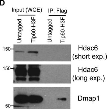
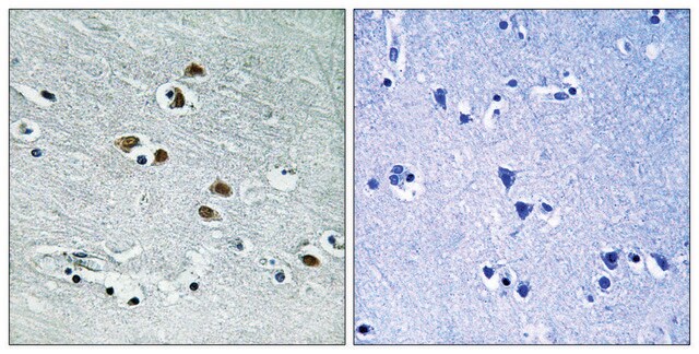
Anti-Histone Deacetylase 6 (HDAC6) recognizes human and mouse HDAC6 (~134 kDa). Applications include immunoblotting, immunoprecipitation, immunocytochemistry and immunohistochemistry.
Applications in which this antibody has been used successfully, and the associated peer-reviewed papers, are given below.
Immunohistochemistry (1 paper)
Western Blotting (1 paper)

Biochem/physiol Actions

Histone Deacetylase 6 (HDAC6) is an enzyme responsible for deacetylation of histones. HDAC6 is a part of transcriptional corepressor complexes. Overexpression or destruction of HDAC6 may lead to Alzheimer, Parkinson and cardiovascular diseases. Overexpression of HDAC6 is implicated in various cancers like malignant melanoma, lung and bladder cancer. It promotes tumorigenesis, cell migration and invasion. HDAC6 also regulates the expression of critical immune system molecules, targets of chemotherapy like programmed death receptor-1 (PD-1). Mutations in HDAC6 causes X-linked chondrodysplasia.

Physical form

Solution in 0.01 M phosphate buffered saline, pH 7.4, containing 15 mM sodium azide.

Disclaimer

Unless otherwise stated in our catalog or other company documentation accompanying the product(s), our products are intended for research use only and are not to be used for any other purpose, which includes but is not limited to, unauthorized commercial uses, in vitro diagnostic uses, ex vivo or in vivo therapeutic uses or any type of consumption or application to humans or animals.

Not finding the right product?  

Try our Product Selector Tool.


Choose from one of the most recent versions:

Certificates of Analysis (COA)

Lot/Batch Number

Don't see the Right Version?

If you require a particular version, you can look up a specific certificate by the Lot or Batch number.

Already Own This Product?

Find documentation for the products that you have recently purchased in the Document Library.

Visit the Document Library

Activation of autophagy in mesenchymal stem cells provides tumor stromal support
Sanchez CG, et al.
Carcinogenesis, 32(7), 964-972 (2011)
Histone deacetylase 6 in cancer
Li T, et al.
Journal of Hematology & Oncology, 11(1), 111-111 (2018)
The human histone deacetylase family
Gray SG and Ekstrom TJ
Experimental Cell Research, 262(2), 75-83 (2001)
The class I-specific HDAC inhibitor MS-275 modulates the differentiation potential of mouse embryonic stem cells
Franci G, et al.
Biology Open, 2(10), 1070-1077 (2013)
Ya Hui et al.
iScience, 27(1), 108689-108689 (2024-01-16)
High glucose has been proved to impair cognitive function in type 2 diabetes, but the underlying mechanisms remain elusive. Here, we found that high glucose increased transcription factors' SP1 O-GlcNAcylation in regulatory T (Treg) cells. Glycosylated SP1 further enhanced HDAC2

Our team of scientists has experience in all areas of research including Life Science, Material Science, Chemical Synthesis, Chromatography, Analytical and many others.

Contact Technical Service