Skip to Content
Merck
All Photos(6)

Key Documents

G8795

Sigma-Aldrich

Anti-GAPDH Antibody

mouse monoclonal, GAPDH-71.1

Synonym(s):

GAPDH Antibody - Monoclonal Anti-GAPDH antibody produced in mouse, Gapdh Antibody, Anti-G3PD, Anti-G3PDH, Anti-Glyceraldehyde-3-phosphate dehydrogenase, Loading Control

Sign Into View Organizational & Contract Pricing


About This Item

MDL number:
UNSPSC Code:
12352203
NACRES:
NA.41
conjugate:
unconjugated
application:
ARR
ELISA (i)
ICC
WB
clone:
GAPDH-71.1, monoclonal
species reactivity:
bovine, turkey, canine, chicken, monkey, mink, mouse, human, rabbit, rat, hamster
citations:
1344
technique(s):
immunocytochemistry: suitable
indirect ELISA: suitable
microarray: suitable
western blot: 0.025-0.05 μg/mL using A431 total cell extract

Product Name

Anti-GAPDH antibody, Mouse monoclonal, clone GAPDH-71.1, purified from hybridoma cell culture

biological source

mouse

Quality Level

conjugate

unconjugated

antibody form

purified from hybridoma cell culture
purified immunoglobulin

antibody product type

primary antibodies

clone

GAPDH-71.1, monoclonal

form

buffered aqueous solution

mol wt

antigen ~37 kDa

species reactivity

bovine, turkey, canine, chicken, monkey, mink, mouse, human, rabbit, rat, hamster

should not react with

prokaryotes

packaging

antibody small pack of 25 μL

concentration

~1 mg/mL

technique(s)

immunocytochemistry: suitable
indirect ELISA: suitable
microarray: suitable
western blot: 0.025-0.05 μg/mL using A431 total cell extract

isotype

IgM

UniProt accession no.

application(s)

research pathology

shipped in

dry ice

storage temp.

−20°C

target post-translational modification

unmodified

Gene Information

human ... GAPDH(2597)
mouse ... Gapdh(14433)
rat ... Gapdh(24383)

General description

Monoclonal Anti-GAPDH (mouse IgM isotype) is derived from the hybridoma GAPDH-71.1 produced by the fusion of mouse myeloma cells (NS1 cells) and splenocytes from BALB/c mice immunized with rabbit GAPDH (glyceraldehyde-3-phosphate dehydrogenase). The gene glyceraldehyde-3-phosphate dehydrogenase (GAPDH) is mapped to human chromosome 12p13. The protein localizes in the cytoplasm but can be translocated to the nucleus depending on cellular conditions.

Specificity

Monoclonal Anti-GAPDH recognizes human, monkey, bovine, canine, rat, mouse, hamster, mink, rabbit, chicken, and turkey GAPDH. It does not cross-react with non-vertebrate and prokaryotic species.

Immunogen

Rabbit GAPDH.

Application

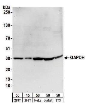
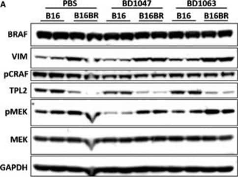
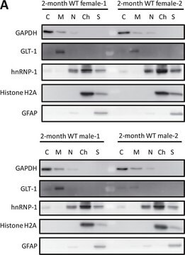
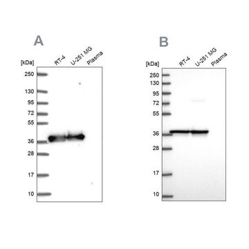
Monoclonal Anti-GAPDH antibody produced in mouse is suitable for western blotting using:
  • protein extracted from heart tissue of mice at a working dilution of 1:25,000
  • myelin and axogliasomal fractions from human CNS
  • nuclear and cytoplasmic fractions from TBP-13Q and TBP-105Q PC12 cells following recovery from heat shock
  • protein from bovine immortalized luteal endothelial cells
  • renal tubular epithelial cell extract
  • proteins from mouse embryonic fibroblasts
  • protein extract from ventricular myocardium tissues
  • A431 total cell extract at a working concentration of 0.025-0.05μg/mL
It is also suitable for immunostaining using leiomyomas and leiomyosarcomas. The antibody can also be used for immunocytochemistry, indirect ELISA and microarray.

Biochem/physiol Actions

Glyceraldehyde-3-phosphate dehydrogenase (GAPDH) is a tetramer containing identical chains. It catalyzes the reversible oxidative phosphorylation of glyceraldehyde-phosphate, which is a critical energy-yielding step in carbohydrate metabolism. It binds to several proteins including actin, tubulin, amyloid precursor, polyglutamine peptides, DRPLA (dentatorubral-pallidoluysian atrophy), and huntingtin. Protein kinase Cι/λbinds and phosphorylates GAPDH. Phosphorylated GAPDH associates with cytoskeletal elements and controls microtubule dynamics in the early secretory pathway. Poly(ADP-ribose) polymerase-1 (PARP1) interacts with GAPDH and thereby mediates brain damage in the presence of oxidative/nitrosative stress. GAPDH forms a part of OCA-S, the multicomponent OCT1 (octamer-motif-binding factor) coactivator complex, which is involved in the S phase-dependent histone H2B transcription. This association is responsible for linking H2B transcriptional machinery to cell cycle regulation and to the cellular metabolic state. GAPDH is also a component of the functional GAIT (interferon-γ-activated inhibitor of translation) mRNP (messenger ribonucleoprotein). GAPDH expression is dysregulated during melanoma progression.

Physical form

Solution in 0.01 M phosphate buffered saline, pH 7.4, containing 15 mM sodium azide.

Storage and Stability

For continuous use, store at 2-8 °C for up to one month.
For extended storage, freeze in working aliquots. Repeated freezing and thawing, or storage in “frostfree” freezers, is not recommended. If slight turbidity occurs upon prolonged storage, clarify the solution by centrifugation before use. Working dilution samples should be discarded if not used within 12 hours.

Disclaimer

Unless otherwise stated in our catalog or other company documentation accompanying the product(s), our products are intended for research use only and are not to be used for any other purpose, which includes but is not limited to, unauthorized commercial uses, in vitro diagnostic uses, ex vivo or in vivo therapeutic uses or any type of consumption or application to humans or animals.

Not finding the right product?  

Try our Product Selector Tool.

Storage Class Code

12 - Non Combustible Liquids

WGK

WGK 1

Flash Point(F)

Not applicable

Flash Point(C)

Not applicable

Personal Protective Equipment

dust mask type N95 (US), Eyeshields, Gloves

Choose from one of the most recent versions:

Certificates of Analysis (COA)

Lot/Batch Number

Don't see the Right Version?

If you require a particular version, you can look up a specific certificate by the Lot or Batch number.

Already Own This Product?

Find documentation for the products that you have recently purchased in the Document Library.

Visit the Document Library

Gloria Ravegnini et al.
Modern pathology : an official journal of the United States and Canadian Academy of Pathology, Inc, 26(5), 743-749 (2012-12-12)
Leiomyoma and leiomyosarcoma share morphological features and smooth muscle differentiation, and both arise most frequently within the uterine corpus of middle-aged women. However, they are considered biologically unrelated tumors due to their disparate clinical, cytogenetic, and molecular features. MED12, the
Frontiers in neurology and neuroscience research null
Qingwen Zheng et al.
American journal of cardiovascular disease, 1(3), 214-226 (2011-11-15)
Protein quality control (PQC) senses and repairs misfolded/unfolded proteins and, if the repair fails, degrades the terminally misfolded polypeptides through an intricate collaboration between molecular chaperones and targeted proteolysis. Proteolysis of damaged proteins is performed primarily by the ubiquitin-proteasome system
Xiaofeng Zuo et al.
The Journal of biological chemistry, 286(25), 22469-22477 (2011-05-06)
Primary cilia are found on many epithelial cell types, including renal tubular epithelial cells, where they participate in flow sensing. Disruption of cilia function has been linked to the pathogenesis of polycystic kidney disease. We demonstrated previously that the exocyst
Hang Zhao et al.
Free radical biology & medicine, 49(4), 641-648 (2010-06-01)
Methionine residues in protein can be oxidized by reactive oxygen species to generate methionine sulfoxide. Aerobic organisms have methionine sulfoxide reductases capable of reducing methionine sulfoxide back to methionine. Methionine sulfoxide reductase A acts on the S-epimer of methionine sulfoxide

Our team of scientists has experience in all areas of research including Life Science, Material Science, Chemical Synthesis, Chromatography, Analytical and many others.

Contact Technical Service