56822
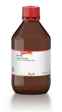
Immersion oil
for microscopy
Synonym(s):
immersion medium for light microscopy
Sign Into View Organizational & Contract Pricing
All Photos(1)
About This Item
Recommended Products
biological source
synthetic
Quality Level
grade
for microscopy
form
liquid
refractive index
n20/D 1.516
viscosity
100-120 mPa.s(20 °C)
density
1.025 g/mL at 20 °C
application(s)
hematology
histology
storage temp.
room temp
Looking for similar products? Visit Product Comparison Guide
General description
Immersion oil is a clear, viscous liquid with optimized refractive properties, specifically modified to closely approximate the refractive index (RI) of glass (ne = 1.5). It is used in conjunction with an objective lens to enhance the resolving power.
Application
Immersion oil is used for high-resolution (1000X) light microscopy work in conjunction with an oil immersion objective lens to optimize microscopic examinations of histological, cytological, hematological, and bacterial specimen material after it has been fixed, embedded, stained, or counterstained, and mounted.
Principle
Immersion oil is applied dropwise to stained and mounted or non-mounted specimen material to form a clear film between the specimen and the microscope lens to eliminate the deflection of incident light and thus substantially enhance the optical efficiency of the lens.
Signal Word
Warning
Hazard Statements
Precautionary Statements
Hazard Classifications
Aquatic Acute 1 - Aquatic Chronic 2
Storage Class Code
10 - Combustible liquids
WGK
WGK 2
Flash Point(F)
Not applicable
Flash Point(C)
Not applicable
Personal Protective Equipment
dust mask type N95 (US), Eyeshields, Gloves
Choose from one of the most recent versions:
Already Own This Product?
Find documentation for the products that you have recently purchased in the Document Library.
Customers Also Viewed
Theis Sommer et al.
Scientific reports, 8(1), 13104-13104 (2018-09-01)
The catalytic mechanism of the cyclic amidohydrolase isatin hydrolase depends on a catalytically active manganese in the substrate-binding pocket. The Mn2+ ion is bound by a motif also present in other metal dependent hydrolases like the bacterial kynurenine formamidase. The
P L Appleton et al.
Journal of microscopy, 234(2), 196-204 (2009-04-29)
Visualizing overall tissue architecture in three dimensions is fundamental for validating and integrating biochemical, cell biological and visual data from less complex systems such as cultured cells. Here, we describe a method to generate high-resolution three-dimensional image data of intact
Thomas P Burghardt et al.
Applied optics, 48(32), 6120-6131 (2009-11-12)
Total internal reflection fluorescence (TIRF) microscopy uses the evanescent field on the aqueous side of a glass/aqueous interface to selectively illuminate fluorophores within approximately 100 nm of the interface. Applications of the method include epi-illumination TIRF, where the exciting light
Our team of scientists has experience in all areas of research including Life Science, Material Science, Chemical Synthesis, Chromatography, Analytical and many others.
Contact Technical Service


