Skip to Content
Merck
All Photos(1)

Key Documents

289R-2

Sigma-Aldrich

Myeloperoxidase (EP151) Rabbit Monoclonal Primary Antibody

Sign Into View Organizational & Contract Pricing


About This Item

UNSPSC Code:
12352203
NACRES:
NA.41

biological source

rabbit

Quality Level

100
500

conjugate

unconjugated

antibody form

culture supernatant

antibody product type

primary antibodies

clone

EP151, monoclonal

description

(For In Vitro Diagnostic Use in Select Regions (See Chart))

form

buffered aqueous solution

species reactivity

human

packaging

bottle of 1.0 mL predilute (289R-27)
bottle of 7.0 mL predilute (289R-28)
vial of 0.1 mL concentrate (289R-24)
vial of 0.5 mL concentrate (289R-25)
vial of 1.0 mL concentrate (289R-26)

manufacturer/tradename

Cell Marque

technique(s)

immunohistochemistry (formalin-fixed, paraffin-embedded sections): 1:100-1:500

isotype

IgG

shipped in

wet ice

storage temp.

2-8°C

Gene Information

human ... MPO(4353)

General description

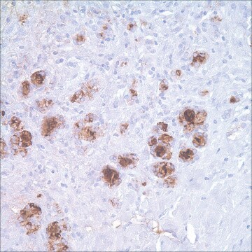
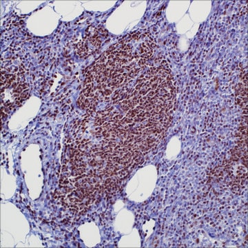
Anti-myeloperoxidase detects granulocytes and monocytes in blood and precursors of granulocytes in the bone marrow. This antibody can detect myeloid cell populations of the bone marrow as well as in other sites.1-8

Quality


IVD

IVD

IVD

RUO

Linkage

Myeloperoxidase Positive Control Slides, Product No. 289S, are available for immunohistochemistry (formalin-fixed, paraffin-embedded sections).

Physical form

Solution in Tris Buffer, pH 7.3-7.7, with 1% BSA and <0.1% Sodium Azide.

Preparation Note

Download the IFU specific to your product lot and formatNote: This requires a keycode which can be found on your packaging or product label.

Other Notes

For Technical Service please contact: 800-665-7284 or email: service@cellmarque.com

Legal Information

Cell Marque is a trademark of Merck KGaA, Darmstadt, Germany

Not finding the right product?  

Try our Product Selector Tool.

Storage Class Code

12 - Non Combustible Liquids

WGK

WGK 2

Flash Point(F)

Not applicable

Flash Point(C)

Not applicable


Certificates of Analysis (COA)

Search for Certificates of Analysis (COA) by entering the products Lot/Batch Number. Lot and Batch Numbers can be found on a product’s label following the words ‘Lot’ or ‘Batch’.

Already Own This Product?

Find documentation for the products that you have recently purchased in the Document Library.

Visit the Document Library

J Hudock et al.
American journal of clinical pathology, 102(1), 55-60 (1994-07-01)
Paraffin sections of granulocytic sarcomas (GS) (n = 30) were immunohistochemically evaluated for CD3, CD15 (LeuM1), CD20 (L26), CD31, CD34, CD43, CD45, CD68 (KP1), lysozyme, myeloperoxidase (BM1), CD45RO (UCHL1), and LN5 with an avidin-biotin amplification system and a peroxidase-based color
G S Pinkus et al.
Modern pathology : an official journal of the United States and Canadian Academy of Pathology, Inc, 4(6), 733-741 (1991-11-01)
Immunohistochemical detection of intracellular myeloperoxidase, a major constituent of primary granules of neutrophilic myeloid cells, was determined in paraffin sections of 161 specimens using a rabbit polyclonal antibody to human myeloperoxidase and an indirect immunoperoxidase technique. In normal tissues and
Z Kaleem et al.
American journal of clinical pathology, 115(6), 876-884 (2001-06-08)
We studied immunophenotypic features of 30 cases of minimally differentiated acute myeloid leukemia (AML-M0) using multiparameter flow cytometry and immunohistochemistry and evaluated the immunophenotypic features of previously reported cases to facilitate correct identification of myeloid lineage. All but 1 of
W H Hamoudi et al.
Archives of pathology & laboratory medicine, 124(2), 315-318 (2000-02-03)
We report a case of primary acute myelomonocytic leukemia involving the bone marrow that resembled sarcomatoid carcinoma. The neoplastic cells in bone marrow biopsy specimens formed cohesive-appearing clusters and cords separated by an immature fibroblastic proliferation and myxoid stroma. Blasts
J Audouin et al.
International journal of surgical pathology, 11(4), 271-282 (2003-11-15)
Extramedullary accumulation of myeloblasts or immature myeloid cells form tumors called myeloid sarcoma in the WHO classification. Such tumors develop in lymphoid organs, bone (skull, orbit, etc.), skin, soft tissue, various mucosae and organs, and the CNS. They may precede

Our team of scientists has experience in all areas of research including Life Science, Material Science, Chemical Synthesis, Chromatography, Analytical and many others.

Contact Technical Service