Skip to Content
Merck
All Photos(2)

Key Documents

MABN153

Sigma-Aldrich

Anti-HuC/HuD Antibody, clone 15A7.1

clone 15A7.1, from mouse

Synonym(s):

ELAV-like protein 3, Hu-antigen C, HuC, Paraneoplastic cerebellar degeneration-associated antigen, Paraneoplastic limbic encephalitis antigen 21, ELAV-like protein 4, Hu-antigen D, HuD, Paraneoplastic encephalomyelitis antigen HuD

Sign Into View Organizational & Contract Pricing


About This Item

UNSPSC Code:
12352203
eCl@ss:
32160702
NACRES:
NA.41

biological source

mouse

Quality Level

antibody form

purified antibody

antibody product type

primary antibodies

clone

15A7.1, monoclonal

species reactivity

human, rat, mouse

technique(s)

immunohistochemistry: suitable (paraffin)
western blot: suitable

isotype

IgG2aκ

NCBI accession no.

UniProt accession no.

shipped in

wet ice

target post-translational modification

unmodified

Gene Information

human ... ELAVL4(1996)

General description

Hu proteins are mammalian embryonic lethal abnormal visual system (ELAV)-like neuronal RNA-binding proteins that contain three RNA recognition motifs. RNA binding protein HuC promotes neuronal development in both the central and the peripheral nervous systems. HuC antibodies are used as markers for developing neurons. HuD may play a role in neuron-specific RNA processing, and be involved in maintaining the proliferative state of cells.

Immunogen

KLH-conjugated linear peptide corresponding to human HuC/HuD.

Application

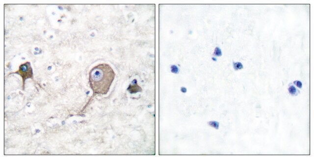
Immunohistochemistry Analysis: A 1:2,000 dilution from a representative lot detected HuC/HuD in human brain and rat hippocampus tissues.
Research Category
Neuroscience
Research Sub Category
Developmental Signaling
This HuC/HuD antibody is validated for use in WB & IHC for the detection of the HuC/HuD protein.

Quality

Evaluated by Western Blotting in human brain tissue lysate.

Western Blotting Analysis: 0.5 µg/mL of this antibody detected HuC/HuD in 200 µg of human brain tissue lysate.

Target description

~ 40 kDa observed. An uncharacterized band may appear at ~45 kDa in some lysates.

Physical form

Format: Purified
Protein G Purified
Purified mouse monoclonal IgG2aκ in buffer containing 0.1 M Tris-Glycine (pH 7.4), 150 mM NaCl with 0.05% sodium azide.

Storage and Stability

Stable for 1 year at 2-8°C from date of receipt.

Analysis Note

Control
Human brain tissue lysate

Other Notes

Concentration: Please refer to the Certificate of Analysis for the lot-specific concentration.

Disclaimer

Unless otherwise stated in our catalog or other company documentation accompanying the product(s), our products are intended for research use only and are not to be used for any other purpose, which includes but is not limited to, unauthorized commercial uses, in vitro diagnostic uses, ex vivo or in vivo therapeutic uses or any type of consumption or application to humans or animals.

Not finding the right product?  

Try our Product Selector Tool.

Storage Class Code

12 - Non Combustible Liquids

WGK

WGK 1

Flash Point(F)

Not applicable

Flash Point(C)

Not applicable


Certificates of Analysis (COA)

Search for Certificates of Analysis (COA) by entering the products Lot/Batch Number. Lot and Batch Numbers can be found on a product’s label following the words ‘Lot’ or ‘Batch’.

Already Own This Product?

Find documentation for the products that you have recently purchased in the Document Library.

Visit the Document Library

Karolína Liška et al.
Neuroendocrinology, 112(4), 384-398 (2021-06-11)
Circadian clocks in the hippocampus (HPC) align memory processing with appropriate time of day. Our study was aimed at ascertaining the specificity of glycogen synthase kinase 3-beta (GSK3β)- and glucocorticoid (GC)-dependent pathways in the entrainment of clocks in individual HPC
Yaxing Gui et al.
Brain pathology (Zurich, Switzerland), 30(1), 151-164 (2019-07-06)
The 18 kDa translocator protein (TSPO) is a widely used target for microglial PET imaging radioligands, but its expression in post-mortem normal and diseased human brain is not well described. We aimed at characterizing the TSPO expression in human control
Joslyn Lay et al.
American journal of physiology. Gastrointestinal and liver physiology, 311(2), G252-G266 (2016-07-02)
The μ-opioid receptor (MOR) is a major regulator of gastrointestinal motility and secretion and mediates opiate-induced bowel dysfunction. Although MOR is of physiological and therapeutic importance to gut function, the cellular and subcellular distribution and regulation of MOR within the
Christine Marques et al.
Acta neuropathologica, 147(1), 56-56 (2024-03-13)
The stimulator of interferon genes (STING) pathway has been implicated in neurodegenerative diseases, including Parkinson's disease and amyotrophic lateral sclerosis (ALS). While prior studies have focused on STING within immune cells, little is known about STING within neurons. Here, we
Clara Muñoz-Castro et al.
Methods in molecular biology (Clifton, N.J.), 2593, 283-305 (2022-12-14)
There is a growing interest in expanding the multiplexing capability of immunohistochemistry to achieve a deeper phenotyping of various cell types in health and disease. Here, we describe a protocol of cyclic multiplex fluorescent immunohistochemistry that enables the labeling of

Our team of scientists has experience in all areas of research including Life Science, Material Science, Chemical Synthesis, Chromatography, Analytical and many others.

Contact Technical Service