Skip to Content
Merck
All Photos(4)

Key Documents

MABC32

Sigma-Aldrich

Anti-p62 (Sequestosome-1) Antibody, clone 11C9.2

clone 11C9.2, from mouse

Synonym(s):

sequestosome 1, EBI3-associated protein of 60 kDa, Paget disease of bone 3, phosphotyrosine independent ligand for the Lck SH2 domain p62, oxidative stress induced like, EBI3-associated protein p60, Phosphotyrosine-independent ligand for the Lck SH2 doma

Sign Into View Organizational & Contract Pricing


About This Item

UNSPSC Code:
12352203
eCl@ss:
32160702
NACRES:
NA.41

biological source

mouse

Quality Level

antibody form

purified antibody

antibody product type

primary antibodies

clone

11C9.2, monoclonal

species reactivity

mouse, rat, human

technique(s)

flow cytometry: suitable
immunocytochemistry: suitable
western blot: suitable

isotype

IgMκ

NCBI accession no.

UniProt accession no.

shipped in

wet ice

target post-translational modification

unmodified

Gene Information

human ... SQSTM1(8878)

General description

p62 is also known as sequestosome-1, and is a 62 kDa ras-GAP associated protein. p62 is thought to bind to src-family tyrosine kinases and may also provide a linker action for activated src-family kinases with their downstream effectors. p62 has an RNA-binding region and may have RNA-binding ability. p62 is associated with activation of transcription factor NF-κB, involved in cell differentiation, immune response and potassium channel regulation. p62 has recently been linked to extrinsic apoptosis and may be a key player in tumorigenesis.

Immunogen

GST-tagged recombinant protein corresponding to human p62 (Sequestosome-1).

Application

Detect p62 (Sequestosome-1) using this Anti-p62 (Sequestosome-1) Antibody, clone 11C9.2 validated for use in WB, FC & IC.
Research Category
Apoptosis & Cancer
Research Sub Category
Apoptosis - Additional
Western Blot Analysis: 0.001 µg/mL from a previous lot detected p62 (Sequestosome-1) in 10 µg of PC12 cell lysate.

Flow Cytometry Analysis: 1.0 µg from a previous lot detected p62 (Sequestosome-1) in the staining of fixed and permeabilized HeLa cells.

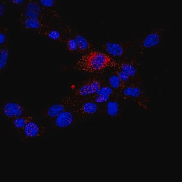
Immunocytochemistry Analysis: A 1:500 dilution from a previous lot detected p62 (Sequestosome-1) in NIH/3T3, A431, and HeLa cells.

Quality

Evaluated by Western Blot in A431 cell lysate.

Western Blot Analysis: 0.001 µg/mL of this antibody detected p62 (Sequestosome-1) in 10 µg of A431 cell lysate.

Target description

~47-60 kDa observed.
The calculated molecular weight of this protein is 47 kDa and also has an isoform at 38 kDa Due to modifications, this protein may be observed up to ~60 kDa in some lysates.

Physical form

Format: Purified
Purified mouse monoclonal IgMκ in buffer containing PBS with 0.05% sodium azide.

Storage and Stability

Stable for 1 year at 2-8°C from date of receipt.

Analysis Note

Control
A431 cell lysate

Disclaimer

Unless otherwise stated in our catalog or other company documentation accompanying the product(s), our products are intended for research use only and are not to be used for any other purpose, which includes but is not limited to, unauthorized commercial uses, in vitro diagnostic uses, ex vivo or in vivo therapeutic uses or any type of consumption or application to humans or animals.

Not finding the right product?  

Try our Product Selector Tool.

Storage Class Code

12 - Non Combustible Liquids

WGK

WGK 2

Flash Point(F)

Not applicable

Flash Point(C)

Not applicable


Certificates of Analysis (COA)

Search for Certificates of Analysis (COA) by entering the products Lot/Batch Number. Lot and Batch Numbers can be found on a product’s label following the words ‘Lot’ or ‘Batch’.

Already Own This Product?

Find documentation for the products that you have recently purchased in the Document Library.

Visit the Document Library

Yue-Mei Feng et al.
Autophagy, 9(9), 1395-1406 (2013-06-27)
Drug addiction is a chronic brain disease that is a serious social problem and causes enormous financial burden. Because mitochondrial abnormalities have been associated with opiate addiction, we examined the effect of morphine on mtDNA levels in rat and mouse
Barbara Jerič et al.
Biochimica et biophysica acta, 1833(10), 2254-2266 (2013-05-21)
The contribution of individual cysteine cathepsins as positive mediators of programmed cell death is dependent on several factors, such as the type of stimuli, intensity and duration of the stimulus, and cell type involved. Of the eleven human cysteine cathepsins
Kleiton Augusto Santos Silva et al.
American journal of physiology. Heart and circulatory physiology, 319(5), H1036-H1043 (2020-09-19)
The small heat shock protein 20 (HSPB6) emerges as a potential upstream mediator of autophagy. Although autophagy is linked to several clinical disorders, how HSPB6 and autophagy are regulated in the setting of heart failure (HF) remains unknown. The goal
The regulatory activity of autophagy in conjunctival fibroblasts and its possible role in vernal keratoconjunctivitis.
Paola Brun et al.
The Journal of allergy and clinical immunology, 146(5), 1210-1213 (2020-04-10)
Huimin Yan et al.
Journal of rheumatic diseases and treatment, 1(1), 5-5 (2015-06-30)
Autophagy plays a central role in various disease processes. However, its contribution to inflammatory arthritides such as rheumatoid arthritis (RA) is unclear. We observed that autophagy is engaged in the K/BxN serum transfer model of RA but autophagic flux is

Our team of scientists has experience in all areas of research including Life Science, Material Science, Chemical Synthesis, Chromatography, Analytical and many others.

Contact Technical Service