MAB1398Z
Anti-PECAM-1 Antibody, clone 2H8, Azide Free
clone 2H8, Chemicon®, from hamster(Armenian)
Synonym(s):
CD31, PECAM-1, Platelet endothelial cell adhesion molecule
About This Item
Recommended Products
biological source
hamster (Armenian)
Quality Level
antibody form
purified immunoglobulin
antibody product type
primary antibodies
clone
2H8, monoclonal
species reactivity
mouse, canine
manufacturer/tradename
Chemicon®
technique(s)
electron microscopy: suitable
flow cytometry: suitable
immunocytochemistry: suitable
immunofluorescence: suitable
immunohistochemistry: suitable
immunoprecipitation (IP): suitable
neutralization: suitable
isotype
IgG
NCBI accession no.
UniProt accession no.
shipped in
dry ice
target post-translational modification
unmodified
Gene Information
dog ... Pecam1(610775)
mouse ... Pecam1(18613)
Related Categories
General description
CD31 has been used to measure angiogenesis in association with tumor recurrence. Other studies have also indicated that CD31 and CD34 can be used as markers for myeloid progenitor cells and recognize different subsets of myeloid leukemia infiltrates (granular sarcomas).
Specificity
Immunogen
Application
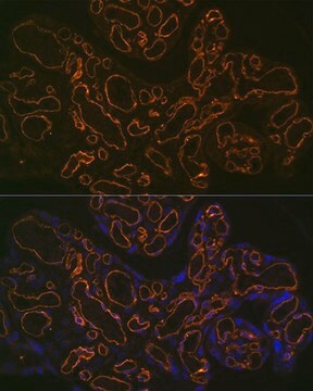
Immunofluorescence Analysis: A representative lot immunostained endothelial cells in formalin-fixed, paraffin-embedded murine peritoneum sections by fluorescent immunohistochemistry (Tanabe, K., et al. (2007). Kidney Int. 71(3):227-238).
Immunocytochemistry Analysis: A representative lot immunostained 4% paraformaldehyde-fixed endothelial cells differentiated from murine ESCs in cultures (Purpura, K.A., et al. (2008). Stem Cells. 26(11):2832-2842).
Immunocytochemistry Analysis: Clone 2H8 immunostained 3% paraformaldehyde-fixed, 0.5% NP-40-permeabilized COS-7 transfectants expressing murine, but not human, CD31 by fluorescent immunocytochemistry (Bogen, S.A., et al. (1992). Am. J. Pathol. 141(4):843-854).
Neutralization Analysis: A representative lot, when injected intravenously in mice, blocked leukocyte emigration in response to thioglycollate injection (Bogen, S., et al. (1994). J. Exp. Med. 179(3):1059-1064).
Neutralization Analysis: A representative lot blocked murine PECAM-mediated aggregation of mouse L cells (Xie, Y., and Muller, W.A. (1993). Proc. Natl. Acad. Sci. U. S. A. 190(12):5569-5573).
Flow Cytometry Analysis: A representative lot detected the expression of exogenously transfected murine CD31 on the surface of mouse L cells (Xie, Y., and Muller, W.A. (1993). Proc. Natl. Acad. Sci. U. S. A. 190(12):5569-5573).
Flow Cytometry Analysis: Clone 2H8 detected CD31 immunoreactivity on the surface of various murine B- and T-cell lines (Bogen, S.A., et al. (1992). Am. J. Pathol. 141(4):843-854).
Immunoprecipitation Analysis: A representative lot immunoprecipitated exogenously expressed murine, but not human, CD31 from COS-7 transfectants (Xie, Y., and Muller, W.A. (1993). Proc. Natl. Acad. Sci. U. S. A. 190(12):5569-5573).
Immunoprecipitation Analysis: Clone 2H8 immunoprecipitated CD31 from murine CTLL T lymphocytes and Sp2/0 B lymphocytes (Bogen, S.A., et al. (1992). Am. J. Pathol. 141(4):843-854).
Electron Microscopy Analysis: Clone 2H8 was biotinylated and immunostained lymphocytes in 0.01% glutaraldehyde-/2% formaldehyde-fxed frozen reactive lymph node tissues from KLH-immunized mice (Bogen, S.A., et al. (1992). Am. J. Pathol. 141(4):843-854).
Immunohistochemistry Analysis: Clone 2H8 immunostained lymphocytes adherent to the sinusoidal and postcapillary venular endothelium of frozen reactive lymph node tissues from KLH-immunized mice (Bogen, S.A., et al. (1992). Am. J. Pathol. 141(4):843-854).
Cell Structure
Adhesion (CAMs)
Linkage
Physical form
Storage and Stability
Analysis Note
Mouse cell lung tissue
Other Notes
Legal Information
Disclaimer
Not finding the right product?
Try our Product Selector Tool.
Storage Class Code
12 - Non Combustible Liquids
WGK
WGK 2
Flash Point(F)
Not applicable
Flash Point(C)
Not applicable
Certificates of Analysis (COA)
Search for Certificates of Analysis (COA) by entering the products Lot/Batch Number. Lot and Batch Numbers can be found on a product’s label following the words ‘Lot’ or ‘Batch’.
Already Own This Product?
Find documentation for the products that you have recently purchased in the Document Library.
Our team of scientists has experience in all areas of research including Life Science, Material Science, Chemical Synthesis, Chromatography, Analytical and many others.
Contact Technical Service


