Skip to Content
Merck
All Photos(2)

Key Documents

AB1930

Sigma-Aldrich

Anti-Integrin alphaV Antibody, CT, Intracellular

serum, Chemicon®

Synonym(s):

CD51

Sign Into View Organizational & Contract Pricing


About This Item

UNSPSC Code:
12352203
eCl@ss:
32160702
NACRES:
NA.41
clone:
polyclonal
application:
ELISA
IHC
IP
RIA
WB
species reactivity:
chicken, hamster, horse, goat, pig, equine, mouse, human, sheep
technique(s):
ELISA: suitable
immunohistochemistry: suitable
immunoprecipitation (IP): suitable
radioimmunoassay: suitable
western blot: suitable
citations:
63

biological source

rabbit

Quality Level

antibody form

serum

antibody product type

primary antibodies

clone

polyclonal

species reactivity

chicken, hamster, horse, goat, pig, equine, mouse, human, sheep

species reactivity (predicted by homology)

rat

manufacturer/tradename

Chemicon®

technique(s)

ELISA: suitable
immunohistochemistry: suitable
immunoprecipitation (IP): suitable
radioimmunoassay: suitable
western blot: suitable

NCBI accession no.

UniProt accession no.

shipped in

wet ice

target post-translational modification

unmodified

Gene Information

human ... ITGAV(3685)

General description

The integrin family of cell adhesion receptors consists of at least 19 membrane-associated heterodimers, composed of an alpha and beta subunit that associate in a non-covalent manner. To date, 16 different alpha-subunits and 8 different beta-subunits have been identified. The structure and functional diversity of the integrin family are based upon the pairing abilities of the individual alpha and beta subunits. Key to these molecular interactions between the integrin receptors and their respective ligands is the recognition of the Arg-Gly-Asp (RGD) sequence, known to be present in the extracellular matrix components fibronectin, vitronectin, collagen, fibrinogen, and von Willebrand factor (Cheresh, 1991). The involvement of integrins in vascular proliferation, adhesion, and wound repair has been well documented. Integrin alphaV is a type I membrane glycoptroein with no I (inserted) domain. The alphaV protein is post-translationally cleaved into a heavy and light chain, which is expressed on the cell surface as a disulfide linked polypeptide.

Specificity

Integrin alphaV. Specificity determined by immunoprecipitation from a detergent extract of 35S methonine labeled endothelial cells. No cross-reactivity to alpha1, alpha2, alpha3, alpha4, or alpha6 integrin subunits was detected. The mature alphaV protein (150 kDa) is composed of a disulfide bonded heavy (125 kDa) and a light (25 kDa) chain derived from a proteolytically cleaved precursor (Suzuki, 1987). The epitope recognized by the antibody is located on the COOH terminal portion of the light chain. By western blot under reducing conditions the antibody detects two major bands at approximately 25 and 27 kDa corresponding to the light chain. The slight differences in sizes of the light chains is thought to be caused by proteolytic cleavage at two alternative sites on the precursor. Similar sites have been reported for alpha6, alpha3, and alphalIbeta (Hogervorst, 1991).

Immunogen

Epitope: C-terminus, intracellular
Synthetic peptide derived from the COOH terminal sequence (cytoplasmic domain) of the human alphaV integrin subunit (SwissProt Accession P06756, amino acids 1022-1034).

Application

Anti-Integrin alphaV Antibody, C-terminus, Intracellular detects level of Integrin alphaV & has been published & validated for use in ELISA, IH, IP, RIA & WB.
ELISA/RIA:
A 1:1,000 dilution of a previous lot was used in ELISA/RIA.

Immunoprecipitation:
5 μL of a previous lot of this antibody is sufficient to precipitate alphaV/beta1 from 5x10⁶ cells.

Immunohistochemistry:
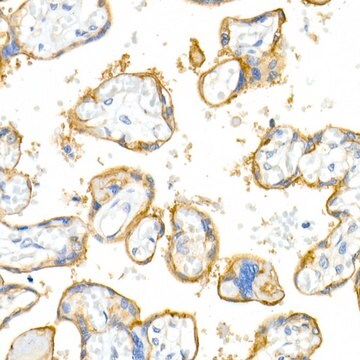
1:1000 dilution from a previous lot was used in immunohistochemistry. (Suggested for use an acetone fixed tissue only.)

Western blot: 1:5000.
The antibody recognizes a single band at approximately 150 kDa when denatured, but not reduced (Hirsch, 1994). Under reducing conditions this antibody recognizes two major bands of 25 and 27 kDa and, to a lesser extent, the full length 150 kDa band.

Optimal working dilutions must be determined by end user.
Research Category
Cell Structure
Research Sub Category
Integrins

Quality

Routinely evaluated by Western Blot on C6 lysates.
Western Blot Analysis: 1:500 dilution of this lot detected INTEGRIN AV light chain on 10 μg of C6 lysates.

Target description

130 kDa; 25 and 27 kDa (reducing conditions)

Physical form

Neat rabbit antiserum in liquid with 0.05% sodium azide.
Unpurified

Storage and Stability

Stable for 1 year at -20°C in undiluted aliquots from date of receipt.

Handling Recommendations:
Upon receipt, and prior to removing the cap, centrifuge the vial and gently mix the solution. Aliquot into microcentrifuge tubes and store at -20°C. Avoid repeated freeze/thaw cycles, which may damage IgG and affect product performance.

Analysis Note

Control
HT-29 colon carcinoma cells

C6 lysates.

Other Notes

Concentration: Please refer to the Certificate of Analysis for the lot-specific concentration.

Legal Information

CHEMICON is a registered trademark of Merck KGaA, Darmstadt, Germany

Disclaimer

Unless otherwise stated in our catalog or other company documentation accompanying the product(s), our products are intended for research use only and are not to be used for any other purpose, which includes but is not limited to, unauthorized commercial uses, in vitro diagnostic uses, ex vivo or in vivo therapeutic uses or any type of consumption or application to humans or animals.

Not finding the right product?  

Try our Product Selector Tool.

Storage Class Code

10 - Combustible liquids

WGK

WGK 1


Certificates of Analysis (COA)

Search for Certificates of Analysis (COA) by entering the products Lot/Batch Number. Lot and Batch Numbers can be found on a product’s label following the words ‘Lot’ or ‘Batch’.

Already Own This Product?

Find documentation for the products that you have recently purchased in the Document Library.

Visit the Document Library

Small molecule proprotein convertase inhibitors for inhibition of embryo implantation.
Ho, H; Singh, H; Heng, S; Nero, TL; Paule, S; Parker, MW; Johnson, AT; Jiao, GS; Nie, G
Testing null
Yoshiyuki Kobayashi et al.
Molecular vision, 22, 436-445 (2016-05-18)
We previously demonstrated that tenascin-C was highly expressed in the fibrovascular membranes (FVMs) of patients with proliferative diabetic retinopathy (PDR). However, its role in the pathogenesis of FVMs has not been determined. The purpose of this study was to investigate
Collagen VI is a basement membrane component that regulates epithelial cell-fibronectin interactions.
Groulx, JF; Gagne, D; Benoit, YD; Martel, D; Basora, N; Beaulieu, JF
Matrix Biology null
Vineet Gupta et al.
Journal of immunology (Baltimore, Md. : 1950), 180(3), 1713-1718 (2008-01-23)
Formation of the integrin alphabeta heterodimer is essential for cell surface expression and function. At the core of the alphabeta interface is a conserved Arg/Lys "finger" from the beta-subunit that inserts into a cup-like "cage" formed of two layers of
Mary Ann Stepp et al.
Experimental cell research, 316(14), 2322-2339 (2010-06-29)
Corneal scarring is a major cause of blindness worldwide and can result from the deposition of abnormal amounts of collagen fibers lacking the correct size and spacing required to produce a clear cornea. Collagen fiber formation requires a preformed fibronectin

Our team of scientists has experience in all areas of research including Life Science, Material Science, Chemical Synthesis, Chromatography, Analytical and many others.

Contact Technical Service