Skip to Content
Merck
All Photos(3)

Key Documents

07-1464

Sigma-Aldrich

Anti-Rac1 Antibody

from rabbit

Synonym(s):

Ras-related C3 Botulinum Toxin Substrate 1, p21-Rac1, Ras-like Protein TC25

Sign Into View Organizational & Contract Pricing


About This Item

UNSPSC Code:
12352203
eCl@ss:
32160702
NACRES:
NA.41
clone:
polyclonal
application:
WB
species reactivity:
human, rat, mouse
technique(s):
western blot: suitable
citations:
6

biological source

rabbit

Quality Level

antibody form

purified antibody

antibody product type

primary antibodies

clone

polyclonal

species reactivity

human, rat, mouse

technique(s)

western blot: suitable

isotype

IgG

NCBI accession no.

UniProt accession no.

shipped in

wet ice

target post-translational modification

unmodified

Gene Information

human ... RAC1(5879)

General description

Rac is a member of the Rho family of small GTP-binding proteins, and plays roles in cytoskeletal actin organization as well as transformation. Rac is activated downstream of cdc42, or independently by Ras signaling, and in turn Rac stimulates Rho. Activation of Rac results in the formation of lamellipodia, whereas cdc42 stimulates filopodia formation, and Rho stimulates formation of actin stress fibers. Effectors of the Rac pathway include NADPH Oxidase and the SAPK1/JNK family of kinases. The latter pathway is activated by the PAK kinases, which are direct targets of Rac.

Specificity

Recognizes Rac1, does not cross react with Cdc42. Rac1 is a member of the Rho family of small GTPases. Small GTPases act as molecular switches in regulating a variety of biological response pathways by cycling from the GDP-bound (inactive) and GTP-bound (active) states. Rac regulates actin polymerization to form lamellipodia.

Immunogen

Synthetic peptide corresponding to amino acids 130-141 of human Rac1 (KEKKLTPITYPQ-C).

Application

Anti-Rac1 Antibody is an antibody against Rac1 for use in WB.
Optimal working dilutions must be determined by end user.
Research Category
Signaling
Research Sub Category
Cytoskeletal Signaling

Quality

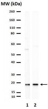
Routinely evaluated by Western Blot on L6 lysates.

Western Blot Analysis: 1:500 dilution of this lot detected RAC1 on 10 μg of L6 lysates.

Target description

21 kDa

Linkage

Replaces: AB4202

Physical form

Format: Purified
Protein A purified
Purified rabbit polyclonal IgG in 0.02 M PBS with 0.15 M NaCl, pH 7.6 and 0.1% sodium azide as a preservative

Storage and Stability

Stable at 2-8°C in undiluted aliquots for up to 6 months from date of receipt.

Analysis Note

Control
Immunoblot/Immunoprecipitation: L6 Cell Lysate or Rat brain microsomal protein preparation
Immunohistochemistry: Rat brain sections

Other Notes

Concentration: Please refer to the Certificate of Analysis for the lot-specific concentration.

Disclaimer

Unless otherwise stated in our catalog or other company documentation accompanying the product(s), our products are intended for research use only and are not to be used for any other purpose, which includes but is not limited to, unauthorized commercial uses, in vitro diagnostic uses, ex vivo or in vivo therapeutic uses or any type of consumption or application to humans or animals.

Not finding the right product?  

Try our Product Selector Tool.

Storage Class Code

10 - Combustible liquids

WGK

WGK 2

Flash Point(F)

Not applicable

Flash Point(C)

Not applicable


Certificates of Analysis (COA)

Search for Certificates of Analysis (COA) by entering the products Lot/Batch Number. Lot and Batch Numbers can be found on a product’s label following the words ‘Lot’ or ‘Batch’.

Already Own This Product?

Find documentation for the products that you have recently purchased in the Document Library.

Visit the Document Library

Joon-Il Jun et al.
Nature cell biology, 12(7), 676-685 (2010-06-08)
Cellular senescence is a recognized mechanism of tumour suppression; however, its contribution to other pathologies is not well understood. We show that the matricellular protein CCN1 (also known as CYR61; cysteine-rich protein 61), which is dynamically expressed at sites of
Jeffrey M Hord et al.
Experimental gerontology, 83, 148-157 (2016-08-19)
Age-related loss of skeletal muscle mass and function, referred to as sarcopenia, is mitigated by lifelong calorie restriction as well as exercise. In aged skeletal muscle fibers there is compromised integrity of the cell membrane that may contribute to sarcopenia.
D Araiza-Olivera et al.
Oncogene, 37(7), 944-952 (2017-10-24)
Activating mutations in the RAC1 gene have recently been discovered as driver events in malignant melanoma. Expression of this gene is associated with melanocyte proliferation, and melanoma cells bearing this mutation are insensitive to BRAF inhibitors such as vemurafenib and
Jeffrey M Hord et al.
Physiological reports, 9(1), e14606-e14606 (2021-01-06)
Reduced mechanical loading results in atrophy of skeletal muscle fibers. Increased reactive oxygen species (ROS) are causal in sarcolemmal dislocation of nNOS and FoxO3a activation. The Nox2 isoform of NADPH oxidase and mitochondria release ROS during disuse in skeletal muscle.
Namrita Kaur et al.
iScience, 25(3), 103973-103973 (2022-03-15)
Myocardial inflammation contributes to cardiomyopathy in diabetic patients through incompletely defined underlying mechanisms. In both human and time-course experimental samples, diabetic hearts exhibited abnormal ER, with a maladaptive shift over time in rodents. Furthermore, as a cardiac ER dysfunction model

Our team of scientists has experience in all areas of research including Life Science, Material Science, Chemical Synthesis, Chromatography, Analytical and many others.

Contact Technical Service