Skip to Content
Merck
All Photos(1)

Key Documents

05-636-AF647

Sigma-Aldrich

Anti-phospho Histone H2A.X (Ser139) Antibody, clone JBW301, Alexa Fluor 647

clone JBW301, 0.5 mg/mL, from mouse

Synonym(s):

Histone H2AX, H2a/x, Histone H2A.X

Sign Into View Organizational & Contract Pricing


About This Item

UNSPSC Code:
12352203
eCl@ss:
32160702
NACRES:
NA.41
conjugate:
ALEXA FLUOR 647
application:
ICC
clone:
JBW301, monoclonal
species reactivity:
human
citations:
13
technique(s):
immunocytochemistry: suitable

biological source

mouse

Quality Level

conjugate

ALEXA FLUOR 647

antibody form

purified antibody

antibody product type

primary antibodies

clone

JBW301, monoclonal

species reactivity

human

species reactivity (predicted by homology)

vertebrates (based on 100% sequence homology)

concentration

0.5 mg/mL

technique(s)

immunocytochemistry: suitable

isotype

IgG1

NCBI accession no.

UniProt accession no.

shipped in

wet ice

target post-translational modification

phosphorylation (pSer139)

Gene Information

human ... H2AX(3014)

General description

As a member of the histone H2A family, histone H2A.x (H2A.x) is a variant histone H2A which replaces conventional H2A in a subset of nucleosomes. H2A.x is involved in the DNA repair of double-strand breakage (DSB) damage on nuclear DNA. After a double strand DNA break, H2A.x is rapidly phosphorylated at serine 139 by ATM kinase and becomes gamma-H2AFX. This phosphorylation step can extend up to several thousand nucleosomes from the actual site of the DSB and may mark the surrounding chromatin for recruitment of proteins required for DNA damage signaling and repair. As a part of posttranslational modifications during apoptosis caused by severe DNA damage, high expression of phosphorylated H2A.x is considered as an accurate indicator of apoptosis.

Immunogen

Linear peptide corresponding to phospho Histone H2A.X (Ser139).

Application

Research Category
Epigenetics & Nuclear Function
Research Sub Category
Histones
This Anti-phospho Histone H2A.X (Ser139) Antibody, clone JBW301, Alexa Fluor 647 is validated for use in ICC for the detection of phospho Histone H2A.X (Ser139).

Quality

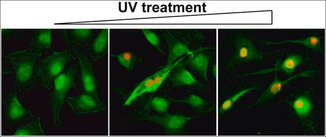
Evaluated by Immunocytochemistry in untreated and staurosporin treated HeLa cells.

Immunocytochemistry Analysis: A 1:100 dilution of this antibody detected phospho Histone H2A.X (Ser139) in staurosporin treated HeLa cells.

Alexa Fluor is a registered trademark of Life Technologies.

Target description

~17 kDa observed. Refer to Cat. No. 05-636 for observed molecular weight information.

Physical form

Protein G Purified
Purified mouse conjugate in buffer containing PBS, 15 mg/ml BSA and 0.1% sodium azide.

Storage and Stability

Stable for 1 year at 2-8°C from date of receipt.

Legal Information

ALEXA FLUOR is a trademark of Life Technologies

Disclaimer

Unless otherwise stated in our catalog or other company documentation accompanying the product(s), our products are intended for research use only and are not to be used for any other purpose, which includes but is not limited to, unauthorized commercial uses, in vitro diagnostic uses, ex vivo or in vivo therapeutic uses or any type of consumption or application to humans or animals.

Not finding the right product?  

Try our Product Selector Tool.

Storage Class Code

12 - Non Combustible Liquids

WGK

WGK 2

Flash Point(F)

Not applicable

Flash Point(C)

Not applicable


Certificates of Analysis (COA)

Search for Certificates of Analysis (COA) by entering the products Lot/Batch Number. Lot and Batch Numbers can be found on a product’s label following the words ‘Lot’ or ‘Batch’.

Already Own This Product?

Find documentation for the products that you have recently purchased in the Document Library.

Visit the Document Library

Yuchen Guo et al.
Genome medicine, 13(1), 166-166 (2021-10-20)
Liver cancer is one of the most commonly diagnosed cancers and the fourth leading cause of cancer-related death worldwide. Broad-spectrum kinase inhibitors like sorafenib and lenvatinib provide only modest survival benefit to patients with hepatocellular carcinoma (HCC). This study aims
Kevin J Lee et al.
Current research in biotechnology, 1, 78-86 (2019-11-01)
Exposures to genotoxic carcinogens and reactive species result in strand breaks and a spectrum of covalent modifications to DNA that can induce mutations and contribute to the initiation and progression of cancer. Measurements of DNA damage within tissue or tumor
Brandon J Aubrey et al.
Genes & development, 32(21-22), 1420-1429 (2018-10-28)
Mutations in Trp53, prevalent in human cancer, are reported to drive tumorigenesis through dominant-negative effects (DNEs) over wild-type TRP53 function as well as neomorphic gain-of-function (GOF) activity. We show that five TRP53 mutants do not accelerate lymphomagenesis on a TRP53-deficient
Mengwen Hu et al.
Nature communications, 13(1), 4510-4510 (2022-08-11)
The ovarian reserve defines the female reproductive lifespan, which in humans spans decades due to robust maintenance of meiotic arrest in oocytes residing in primordial follicles. Epigenetic reprogramming, including DNA demethylation, accompanies meiotic entry, but the chromatin changes that underpin
Akihiko Sakashita et al.
Nature structural & molecular biology, 27(10), 967-977 (2020-09-09)
Gene regulation in the germline ensures the production of high-quality gametes, long-term maintenance of the species and speciation. Male germline transcriptomes undergo dynamic changes after the mitosis-to-meiosis transition and have been subject to evolutionary divergence among mammals. However, the mechanisms

Our team of scientists has experience in all areas of research including Life Science, Material Science, Chemical Synthesis, Chromatography, Analytical and many others.

Contact Technical Service