Skip to Content
Merck
All Photos(2)

Key Documents

F0772

Sigma-Aldrich

Monoclonal Anti-CD8−FITC antibody produced in mouse

clone UCHT-4, purified immunoglobulin, buffered aqueous solution

Synonym(s):

Monoclonal Anti-CD8

Sign Into View Organizational & Contract Pricing


About This Item

UNSPSC Code:
12352203
NACRES:
NA.44

biological source

mouse

Quality Level

conjugate

FITC conjugate

antibody form

purified immunoglobulin

antibody product type

primary antibodies

clone

UCHT-4, monoclonal

form

buffered aqueous solution

species reactivity

human

technique(s)

flow cytometry: 10 μL using 1 × 10⁶ cells

isotype

IgG2a

UniProt accession no.

shipped in

wet ice

storage temp.

2-8°C

target post-translational modification

unmodified

Gene Information

Looking for similar products? Visit Product Comparison Guide

Related Categories

General description

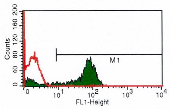
Monoclonal Anti-Human CD8 (mouse IgG2a isotype) is derived from the hybridoma produced by the fusion of mouse myeloma cell line NS-1 and splenocytes from Balb/c mice immunized with human thymocytes followed by peripheral blood T cells. The CD8 antigen is strongly expressed on approximately one-third of mature T cells (cytotoxic/suppressor T cells). Monoclonal Anti-CD8 does not stain B lymphocytes, monocytes, or granulocytes.

Specificity

Recognizes the CD8 human T cytotoxic/suppressor lymphocyte cell surface glycoprotein. Approximately 90% of thymocytes are stained in suspension. In thymus sections, both cortical and medullary thymocytes are stained. A subset of NK cells express this antigen weakly. The epitope recognized by this clone is sensitive to routine formalin fixation and paraffin embedding.
3rd Workshop: code no. 567

Immunogen

human thymocytes followed by peripheral blood T cells.

Application

FITC Monoclonal Anti-Human CD8 may be used for:
  • Immunofluorescent staining,
  • Enumeration of total T cytotoxic/suppressor lymphocytes in bone marrow, blood and other body fluids,
  • Identification and localization of T cytotoxic/ suppressor lymphocytes in lymphoid and other tissues,
  • Analysis of cell mediated cytotoxicity,
  • Studies of immunoregulation in health and disease,
  • Investigation of NK cells, Complement mediated cytolysis of CD8 expressing cells

Biochem/physiol Actions

CD8a (Cluster of Differentiation 8a) is a human gene. CD8 bind to class I MHC and the co-receptors during the process of cytotoxic T-cell activation. T cells that express T cell receptor (TCR) complexes, which lack CD3 δ, exhibit severely impaired positive selection and TCR-mediated activation of CD8 single-positive T cells. CD3 δ couples with TCR. The CD3 associated with CD8 is required for effective activation and positive selection of CD8+ T cells. CD8 expression is a recurrent rare phenomenon in patients with chronic lymphocytic leukemia (CLL).

Target description

CD8 is a two chain complex (α/α, α/β) receptor for Class I MHC molecules. The receptor′s cytoplasmic portion is associated with p56lck protein tyrosine kinase which phosphorylates nearby proteins. The antigen is strongly expressed on approximately one third of mature T cells (cytotoxic/suppressor T cells).

Physical form

Solution in 0.01 M phosphate buffered saline, pH 7.4, containing 1% bovine serum albumin and 15 mM sodium azide.

Preparation Note

Prepared by conjugation to fluorescein isothiocyanate isomer I (FITC). This green dye is efficiently excited at 495 nm and emits at 525 nm.

Disclaimer

Unless otherwise stated in our catalog or other company documentation accompanying the product(s), our products are intended for research use only and are not to be used for any other purpose, which includes but is not limited to, unauthorized commercial uses, in vitro diagnostic uses, ex vivo or in vivo therapeutic uses or any type of consumption or application to humans or animals.

Not finding the right product?  

Try our Product Selector Tool.

Storage Class Code

10 - Combustible liquids

WGK

nwg

Flash Point(F)

Not applicable

Flash Point(C)

Not applicable


Certificates of Analysis (COA)

Search for Certificates of Analysis (COA) by entering the products Lot/Batch Number. Lot and Batch Numbers can be found on a product’s label following the words ‘Lot’ or ‘Batch’.

Already Own This Product?

Find documentation for the products that you have recently purchased in the Document Library.

Visit the Document Library

CXCL9 promotes prostate cancer progression through inhibition of cytokines from T cells
Tan S, et al.
Molecular Medicine Reports, 18(2), 1305-1310 (2018)
G F Gao et al.
Immunology today, 21(12), 630-636 (2000-12-15)
In recent years, substantial progress has been made towards understanding the molecular basis for CD8 binding to class I MHC and the coreceptor's role in cytotoxic T-cell activation. Here, we review the structural, mechanistic and functional studies that point to
Christian Schütz et al.
Blood, 111(7), 3546-3552 (2007-12-22)
Several cell-based immunotherapy strategies have been developed to specifically modulate T cell-mediated immune responses. These methods frequently rely on the utilization of tolerogenic cell-based antigen-presenting cells (APCs). However, APCs are highly sensitive to cytotoxic T-cell responses, thus limiting their therapeutic
C L Kohem et al.
Arthritis and rheumatism, 39(5), 844-854 (1996-05-01)
To delineate in greater detail the phenotype of T cells that reside in the synovial tissue (ST) and synovial fluid (SF) of patients with rheumatoid arthritis (RA), in order to determine their precise differentiation status, and to determine whether the
Wolfgang Kern et al.
Cytometry. Part B, Clinical cytometry, 82(3), 145-150 (2011-12-07)
In patients with chronic lymphocytic leukemia (CLL), aberrant expression of the T-lineage antigen CD8 was reported in low frequencies. The clinical impact of this phenomenon remains in discussion. We analyzed 5,523 patients with CLL (21-97 years) by multiparametric flow cytometry

Our team of scientists has experience in all areas of research including Life Science, Material Science, Chemical Synthesis, Chromatography, Analytical and many others.

Contact Technical Service