Skip to Content
Merck
All Photos(2)

Documents

C2931

Sigma-Aldrich

Monoclonal Anti-Cytokeratin, pan antibody produced in mouse

clone C-11, ascites fluid

Synonym(s):

Monoclonal Anti-Pan Cytokeratin

Sign Into View Organizational & Contract Pricing


About This Item

UNSPSC Code:
12352203
NACRES:
NA.41

biological source

mouse

Quality Level

conjugate

unconjugated

antibody form

ascites fluid

antibody product type

primary antibodies

clone

C-11, monoclonal

contains

15 mM sodium azide

species reactivity

bovine, mouse, frog, human, kangaroo rat, rat

technique(s)

immunohistochemistry (formalin-fixed, paraffin-embedded sections): suitable using protease-digested sections of human or animal tissues
immunohistochemistry (frozen sections): suitable
indirect immunofluorescence: 1:400 using protease-digested, formalin-fixed, paraffin-embedded sections of human or animal tissues
western blot: suitable

isotype

IgG1

shipped in

dry ice

storage temp.

−20°C

target post-translational modification

unmodified

Looking for similar products? Visit Product Comparison Guide

General description

Intermediate-sized filaments are abundant cytoplasmic structural proteins in most vertebrate cells. Cytokeratins, a group of at least 29 different proteins, are characteristic of epithelial and trichocytic cells. Cytokeratins 4, 5, 6 and 8 are members of the type II neutral-to-basic subfamily. Cytokeratin peptide 4 (59 kDa) is the secondary type II keratin expressed in noncornified stratified squamous epithelia. Cytokeratin peptide 5 (58 kDa) is the primary type II keratin in stratified epithelia, while cytokeratin type 8 (52 kDa) is a major type II keratin in simple epithelia. Cytokeratin 6 (56 kDa) is a "hyperproliferation" cytokeratin expressed in tissues with natural or pathological high turnover. Cytokeratins 10, 13 and 18 are members of the type I acidic subfamily. Cytokeratin peptide 10 (56 kDa) is the secondary type I keratin expressed in cornified epithelia. Cytokeratin 13 (54 kDa) is the secondary type I keratin expressed in non-cornified stratified squamous epithelia. Cytokeratin 18 (45 kDa) is the primary type I keratin expressed in simple epithelial cells.
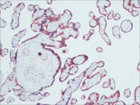
Monoclonal Anti-Pan Cytokeratin (clone C-11) recognizes human cytokeratins 4, 5, 6, 8, 10, 13 and 18 in immunoblotting. The antibody reacts with simple, cornifying and non-cornifying squamous epithelia and pseudostratified epithelia. It does not react with non-epithelial normal human tissues. This antibody can be applied to methanol or acetone-fixed, frozen sections, and to formalin-fixed, paraffin-embedded human tissues. Increased staining intensity is seen following proteolytic treatment of formalin fixed tissue. Similarly, methacarn-fixed material is also suitable for cytokeratin demonstration. Monoclonal Anti-Pan cytokeratin exhibits a wide interspecies cross-reactivity (e.g., human, bovine, rat, frog).

Immunogen

keratin-enriched preparation from cultured human epidermoid carcinoma cell line A431.

Application

Monoclonal Anti-Cytokeratin, pan antibody has been used in immunofluorescence microscopy.
Monoclonal anti-cytokeratins are specific markers of epithelial cell differentiation and have been widely used as tools in tumor identification and classification. Mouse monoclonal clone C-11 anti-cytokeratin, pan antibody is a broad spectrum antibody which recognizes an epitope present in most human epithelial tissues. It facilitates typing of normal, metaplastic and neoplastic cells. It may aid in the discrimination of carcinomas and non-epithelial tumors such as sarcomas, lymphomas and neural tumors. It is also useful in detecting micrometastases in lymph nodes, bone marrow and other tissues, and for determining the origin of poorly differentiated tumors. It is useful for staining of cultured epithelial cell lines. Mouse monoclonal clone C-11 anti-Cytokeratin, pan antibody may be used for the localization of cytokeratins using various immunochemical assays such as immunoblotting, dot blotting and immunohistochemistry (immunofluorescence and immunoenzymatic staining).

Biochem/physiol Actions

Cytokeratins are usually used to determine epithelial differentiation. They help to maintain the integrity of the epithelium of the anterior segment of the eye.

Disclaimer

Unless otherwise stated in our catalog or other company documentation accompanying the product(s), our products are intended for research use only and are not to be used for any other purpose, which includes but is not limited to, unauthorized commercial uses, in vitro diagnostic uses, ex vivo or in vivo therapeutic uses or any type of consumption or application to humans or animals.

Not finding the right product?  

Try our Product Selector Tool.

Storage Class Code

10 - Combustible liquids

WGK

nwg

Flash Point(F)

Not applicable

Flash Point(C)

Not applicable


Certificates of Analysis (COA)

Search for Certificates of Analysis (COA) by entering the products Lot/Batch Number. Lot and Batch Numbers can be found on a product’s label following the words ‘Lot’ or ‘Batch’.

Already Own This Product?

Find documentation for the products that you have recently purchased in the Document Library.

Visit the Document Library

Non-Ocular Sources for Cell-Based Ocular Surface Reconstruction
Ocular Surface Disease: Cornea, Conjunctiva and Tear Film, 373-383 (2013)
Variation in composition of the intervillous space lining in term placentas of mothers with pre-eclampsia
Hottor B, et al.
Placenta, 31(5), 409-417 (2010)
Tao Li et al.
Oncology letters, 12(2), 1129-1131 (2016-07-23)
A 49-year-old female patient presented with a massive left renal tumor, recurrent left flank pain and gross hematuria. The tumor was accompanied by a renal vein tumor thrombus, pancreatic infiltration and a solitary adrenal metastasis. Radical nephrectomy, distal pancreatectomy, ipsilateral
Sara Hägglund et al.
Transboundary and emerging diseases, 67(1), 133-148 (2019-08-17)
Foot-and-mouth disease virus (FMDV) causes a highly contagious vesicular disease in livestock, with serious consequences for international trade. The virus persists in the nasopharynx of cattle and this slows down the process to obtain an FMDV-free status after an outbreak.
Md Rashedul Islam et al.
The Journal of reproduction and development, 62(3), 271-278 (2016-03-08)
Endometrial modulation is essential for the preservation of normal uterine physiology, and this modulation is driven by a number of growth factors. The present study investigated the mitogenic, motogenic, and morphogenic effects of epidermal growth factor (EGF) and hepatocyte growth

Our team of scientists has experience in all areas of research including Life Science, Material Science, Chemical Synthesis, Chromatography, Analytical and many others.

Contact Technical Service