Z764000
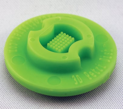
MicroTissues® 3D Petri Dish® micro-mold spheroids
size S, 16 x 16 array, fits 12 well plates
Sinónimos:
3D, 3D Cell Culture
Iniciar sesiónpara Ver la Fijación de precios por contrato y de la organización
About This Item
UNSPSC Code:
41121812
NACRES:
NB.14
Productos recomendados
material
spherical
size
S
sterility
sterile; autoclaved
feature
lid: no
16 x 16 array
packaging
pack of 6 ea
manufacturer/tradename
MicroTissues Inc. 12-256
volume
190 μL
¿Está buscando productos similares? Visita Guía de comparación de productos
General description
Six autoclavable precision micro-molds to cast 3D Petri Dish for forming small spheroids. 3D Petri Dish for use in 12-well plate. Each micro-mold forms 256 circular recesses in a 16 x 16 array.
- Nominal dimensions of each 3D culture recess: diam. 300 μm x D 800 μm
- Micro-molds also form single chamber for cell seeding
When the gelled agarose is removed from the micro-mold, it is transferred to a standard 12 well or 24 well tissue culture dish and equilibrated with cell culture medium.
Since the agarose is transparent, the spheroids or microtissues that form at the bottom of each agarose micro-well can be easily viewed using a standard inverted microscope using phase contrast, bright field or fluorescence microscopy.
Since the agarose is transparent, the spheroids or microtissues that form at the bottom of each agarose micro-well can be easily viewed using a standard inverted microscope using phase contrast, bright field or fluorescence microscopy.
- The micro-molds are reusable up to 12 times and can be sterilized via a standard steam autoclave (30 min, dry cycle)
- The micro-molds should be stored in a covered container to maintain sterility and to avoid collecting dust or fibres on their small features
- Any microscope with sufficient magnification and resolution functions for cell based views or images will be suitable for use with the 3D Petri Dish products
Legal Information
3D Petri Dish is a registered trademark of MicroTissues Inc.
MicroTissues is a registered trademark of MicroTissues Inc.
Elija entre una de las versiones más recientes:
Certificados de análisis (COA)
Lot/Batch Number
Lo sentimos, en este momento no disponemos de COAs para este producto en línea.
Si necesita más asistencia, póngase en contacto con Atención al cliente
¿Ya tiene este producto?
Encuentre la documentación para los productos que ha comprado recientemente en la Biblioteca de documentos.
Los clientes también vieron
Minjae Kim et al.
Journal of cell science, 132(19) (2019-09-08)
Cultured rat primitive extraembryonic endoderm (pXEN) cells easily form free-floating multicellular vesicles de novo, exemplifying a poorly studied type of morphogenesis. Here, we reveal the underlying mechanism and the identity of the vesicles. We resolve the morphogenesis into vacuolization, vesiculation
Nuestro equipo de científicos tiene experiencia en todas las áreas de investigación: Ciencias de la vida, Ciencia de los materiales, Síntesis química, Cromatografía, Analítica y muchas otras.
Póngase en contacto con el Servicio técnico


