Saltar al contenido
Merck

S2532

Sigma-Aldrich

Anti-S100B Antibody

mouse monoclonal, SH-B1

Sinónimos:

Anti-NEF, Anti-S100, Anti-S100-B, Anti-S100beta

Iniciar sesiónpara Ver la Fijación de precios por contrato y de la organización


About This Item

MDL number:
UNSPSC Code:
12352203
NACRES:
NA.41
conjugate:
unconjugated
application:
ARR
DB
ELISA (i)
IHC (p)
RIA
clone:
SH-B1, monoclonal
species reactivity:
feline, pig, bovine, sheep, goat, rabbit, canine, human, rat
citations:
324
technique(s):
dot blot: suitable using denatured-reduced preparations
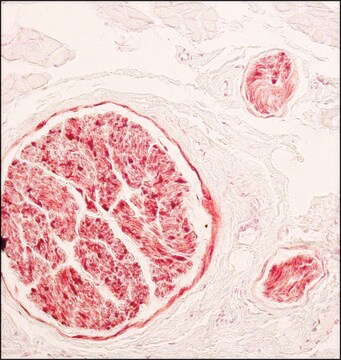
immunohistochemistry (formalin-fixed, paraffin-embedded sections): 1:1000 using protease-digested sections of human tongue
indirect ELISA: suitable
microarray: suitable
radioimmunoassay: suitable

Nombre del producto

Monoclonal Anti-S-100 (β-Subunit) antibody produced in mouse, clone SH-B1, ascites fluid

biological source

mouse

Quality Level

conjugate

unconjugated

antibody form

ascites fluid

antibody product type

primary antibodies

clone

SH-B1, monoclonal

contains

15 mM sodium azide

species reactivity

feline, pig, bovine, sheep, goat, rabbit, canine, human, rat

technique(s)

dot blot: suitable using denatured-reduced preparations
immunohistochemistry (formalin-fixed, paraffin-embedded sections): 1:1000 using protease-digested sections of human tongue
indirect ELISA: suitable
microarray: suitable
radioimmunoassay: suitable

isotype

IgG1

UniProt accession no.

shipped in

dry ice

storage temp.

−20°C

target post-translational modification

unmodified

Gene Information

human ... S100B(6285)
rat ... S100b(25742)

General description

S100 gene is mapped on human chromosome 21q22.3 and codes for an acidic calcium binding protein B, which weighs 21 kDa. The gene is predominantly expressed in astrocytes, glial cells, Schwann cells and also found in adipose cells, skin, glioblastoma multiforme and melanoma. The protein is localized in extracellular space and cytoplasm.
S100B (S-100β Subunit) belongs to the S100 family of calcium-binding proteins. It is expressed in melanocytes.
S-100 is a set of small, thermolabile, highly acidic dimer proteins of approximately 20kDa which are widely distributed in different tissues. Dimeric combinations of two chains, the α-chain (93 amino acids,10.4kDa) and the β-chain (91 amino acids, 10.5kDa), form the three known subtypes of S-100: S-100ao (αα), S-100a (αβ) and S-100b (ββ).
Monoclonal Anti-S-100 (β-subunit) (mouse IgG1 isotype) is derived from the SH-B1 hybridoma produced by the fusion of mouse myeloma cells and splenocytes from an immunized mouse.

Specificity

This antibody recognizes an epitope located on the β chain (i.e. in S-100a and S-100b) but not on the α chain of S-100 (i.e. in S-100a and S-100ao). In ELISA, recognition of S-100 subunit by clone SH-B1 is independent of Ca2+. The product does not react with other members of the EF-hand family such as, calmodulin, parvalbumin, intestinal calcium-binding protein and myosin light chain. In immunohistochemistry, the antibody detects normal and neoplastic S-100 β subunit-containing cells (e.g. Schwann cells, chondrocytes, melanocytes, and melanotic tumors) in protease-digested, formalin-fixed, paraffin-embedded tissues.

Immunogen

Bovine brain S-100b

Application

Monoclonal Anti-S-100 (β-Subunit) antibody produced in mouse has been used in the following:
  • ELISA
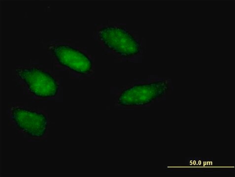
  • Immunofluorescence
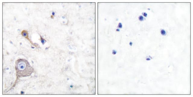
  • Immunohistochemistry
  • Western blotting

Biochem/physiol Actions

Increased expression of S100 is observed in neuronal damage and thus, serves as a biomarker for detecting brain damage and also in several cancers. Also, it is highly permeable through the blood-brain barrier. The gene is known to be associated with cancer progression.
S100β participates in multiple cellular activities including cell growth, cell structure, cell-cell communication, energy metabolism, contraction, and intracellular signal transduction. In utero exposure to cocaine resulting in aberrant S100β levels is linked with neurobehavioral abnormalities and microcephaly. Fetuses with anencephaly show aberrant cell immunoreactivity. Increased levels of this protein are found in plasma of patients with brain damage. S100β is a candidate biochemical marker for spinal cord injury (SCI). Elevated expression of this protein in subpopulations of reactive astrocytes might be linked with hippocampal structural reorganization post-kainic acid (KA)-mediated neurodegeneration.

Physical form

The product is provided as ascites fluid containing 15 mM sodium azide as a preservative.

Storage and Stability

For continuous use, store at 2-8 °C for up to one month. For extended storage, the solution may be frozen in working aliquots. Repeated freezing and thawing is not recommended. Storage in "frost-free" freezers is not recommended. If slight turbidity occurs upon prolonged storage, clarify by centrifugation before use.

Disclaimer

Unless otherwise stated in our catalog or other company documentation accompanying the product(s), our products are intended for research use only and are not to be used for any other purpose, which includes but is not limited to, unauthorized commercial uses, in vitro diagnostic uses, ex vivo or in vivo therapeutic uses or any type of consumption or application to humans or animals.

¿No encuentra el producto adecuado?  

Pruebe nuestro Herramienta de selección de productos.

Storage Class

10 - Combustible liquids

wgk_germany

WGK 3

flash_point_f

Not applicable

flash_point_c

Not applicable


Elija entre una de las versiones más recientes:

Certificados de análisis (COA)

Lot/Batch Number

¿No ve la versión correcta?

Si necesita una versión concreta, puede buscar un certificado específico por el número de lote.

¿Ya tiene este producto?

Encuentre la documentación para los productos que ha comprado recientemente en la Biblioteca de documentos.

Visite la Librería de documentos

Circulating S100beta protein is increased in intrauterine growth-retarded fetuses.
Gazzolo D et al
Pediatric Research, 51(2), 215-219 (2002)
Differential expression of S100beta and glial fibrillary acidic protein in the hippocampus after kainic acid-induced lesions and mossy fiber sprouting in adult rat.
Bendotti C et al
Experimental Neurology, 161(1), 317-329 (2000)
Samantha L Sison et al.
Journal of virology, 93(17) (2019-06-21)
The herpesvirus human cytomegalovirus (HCMV) is a leading cause of congenital birth defects. Infection can result in infants born with a variety of symptoms, including hepatosplenomegaly, microcephaly, and developmental disabilities. Microcephaly is associated with disruptions in the neural progenitor cell
Reprogramming of neural progenitor cells into induced pluripotent stem cells in the absence of exogenous Sox2 expression.
Eminli S et al
Stem Cells, 26(10), 2467-2474 (2008)
S100B is a potential disease activity marker in nonsegmental vitiligo.
Speeckaert R, et al.
The Journal of Investigative Dermatology, 137(7), 1445-1453 (2017)

Nuestro equipo de científicos tiene experiencia en todas las áreas de investigación: Ciencias de la vida, Ciencia de los materiales, Síntesis química, Cromatografía, Analítica y muchas otras.

Póngase en contacto con el Servicio técnico