MABS2283
Anti-PIP2 Antibody, clone KT10
Sinónimos:
Phosphatidylinositol 4;5-bisphosphate;PtdIns(4;5)P2
About This Item
FACS
ICC
inhibition assay
flow cytometry: suitable
immunocytochemistry: suitable
inhibition assay: suitable
Productos recomendados
biological source
mouse
Quality Level
conjugate
unconjugated
antibody form
purified antibody
antibody product type
primary antibodies
clone
KT10, monoclonal
mol wt
calculated mol wt 1.042 kDa
purified by
using Protein A
species reactivity
rat, mouse, human
species reactivity (predicted by homology)
bovine
packaging
antibody small pack of 100 μg
technique(s)
ELISA: suitable
flow cytometry: suitable
immunocytochemistry: suitable
inhibition assay: suitable
isotype
IgG2a
epitope sequence
Unknown
Protein ID accession no.
UniProt accession no.
shipped in
ambient
target post-translational modification
unmodified
Gene Information
mouse ... Stag1(20842)
General description
Specificity
Immunogen
Application
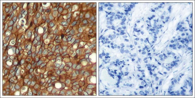
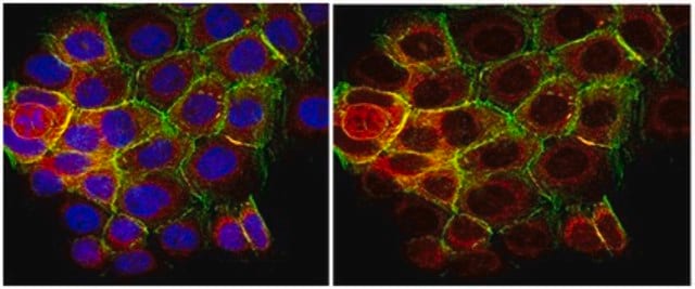
Evaluated by Immunocytochemistry in K562 cells.
Immunocytochemistry Analysis: A 1:100 dilution of this antibody detected PI(4,5)P2 (PIP2) in K562 cells.
Tested Applications
ELISA Analysis: A representative lot detected PIP2 in ELISA applications (Fukami, K. et al. (1988). Proc Natl Acad Sci USA. 85(23): 9057-61).
Immunocytochemistry Analysis: A representative lot detected PIP2 in Immunocytochemistry applications (Yoneda, A., et al. (2020). Biochem Biophys Res Commun. 527(4): 1050-1056).
Flow Cytometry Analysis: Analysis: A representative lot detected PIP2 in Flow Cytometry applications (Yoneda, A., et al. (2020). Biochem Biophys Res Commun. 527(4): 1050-1056).
Inhibition Assay: A representative lot of this antibody inhibited intracellular breakdown of PIP2 and also blocked the proliferation of Src- and erbB-transformed cells. (Fukami, K. et al. (1988). Proc Natl Acad Sci USA. 85(23): 9057-61).
Note: Actual optimal working dilutions must be determined by end user as specimens, and experimental conditions may vary with the end user
Physical form
Storage and Stability
Other Notes
Disclaimer
¿No encuentra el producto adecuado?
Pruebe nuestro Herramienta de selección de productos.
Storage Class
12 - Non Combustible Liquids
wgk_germany
WGK 1
Certificados de análisis (COA)
Busque Certificados de análisis (COA) introduciendo el número de lote del producto. Los números de lote se encuentran en la etiqueta del producto después de las palabras «Lot» o «Batch»
¿Ya tiene este producto?
Encuentre la documentación para los productos que ha comprado recientemente en la Biblioteca de documentos.
Nuestro equipo de científicos tiene experiencia en todas las áreas de investigación: Ciencias de la vida, Ciencia de los materiales, Síntesis química, Cromatografía, Analítica y muchas otras.
Póngase en contacto con el Servicio técnico
![MK571, Sodium Salt A selective, competitive antagonist of leukotriene D4 (LTD4) (Ki = 2.1 nM for inhibition of [³H]LTD4 binding to human lung membranes).](/deepweb/assets/sigmaaldrich/product/images/110/905/114b43f4-ee35-4aba-ab41-09f068177368/640/114b43f4-ee35-4aba-ab41-09f068177368.jpg)
