IgG antibody consists of four subclasses : IgG1, IgG2, IgG3 and IgG4. The IgG structure possesses four polypeptide chains containing two identical γ heavy (H) chains and two identical κ or λ light (L) chains of 50 kDa and 25 kDa respectively. The chains are interlinked with a disulfide bond.
IgG antibody subtype is the most abundant serum immunoglobulins of the immune system. It is secreted by B cells and is found in blood and extracellular fluids and provides protection from infections caused by bacteria, fungi and viruses. Maternal IgG is transferred to fetus through the placenta that is vital for immune defence of the neonate against infections Anti-Mouse IgG (whole molecule)-FITC antibody is specific for mouse IgG. Goat anti-mouse IgG is conjugated to Fluorescein Isothiocyanate (FITC), Isomer I. Following conjugation, unbound FITC is removed by extensive dialysis.
Immunogen
Purified mouse IgG
Application
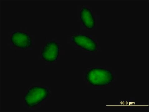
Anti-Mouse IgG (whole molecule)−FITC antibody produced in goat has been used in flow cytometry.
The antibody may be used for immunofluorescent staining of mouse spleen cells at a working dilution of 1:64. A working antibody dilution of 1:40 was used to stain the conidia of Botrytis cinerea for immunofluorescence. The antibody was also used for immunofluorescence of primary luteal cell cultures incubated with primary antibody against CD45 and rhizoids of Chara globularis incubated with anti-human spectrin antibody.
Biochem/physiol Actions
IgG (immunoglobulin G) antibody has its function similar to IgM antibody in complement system activation. IgG participates in hypersensitivity type II and type III. It helps in opsonization, complement fixation and antibody-dependent cell-mediated cytotoxicity.
Physical form
Solution in 0.01 M phosphate buffered saline, pH 7.4, containing 15 mM sodium azide.
Disclaimer
Unless otherwise stated in our catalog or other company documentation accompanying the product(s), our products are intended for research use only and are not to be used for any other purpose, which includes but is not limited to, unauthorized commercial uses, in vitro diagnostic uses, ex vivo or in vivo therapeutic uses or any type of consumption or application to humans or animals.
There is a strong need for rapid and reliable epitope mapping methods that can keep pace with the isolation of increasingly larger numbers of mAbs. We describe here the identification of a conformational epitope using Phage-based Representation OF ImmunoLigand Epitope
Development of a monoclonal antibody-based immunodetection assay for Botrytis cinerea
The journal of histochemistry and cytochemistry : official journal of the Histochemistry Society, 37(7), 1013-1024 (1989-07-01)
Rat monoclonal antibodies (MAb) directed to mouse Ig heavy and light chain determinants were produced. A rat anti-mouse light chain MAb (RAMOL-1) which bound to all (24/24) mouse Ig of the kappa light chain type and with varying strength to
Spectrin-like epitopes were immunochemically detected and immunofluorescently localized in gravitropically tip-growing rhizoids and protonemata of characean algae. Antiserum against spectrin from chicken erythrocytes showed cross-reactivity with rhizoid proteins at molecular masses of about 170 and 195 kD. Confocal microscopy revealed
International journal of molecular medicine, 40(2), 474-482 (2017-06-29)
The pathogenesis of Japanese encephalitis virus (JEV) is complex and unclearly defined, and in particular, the effects of the JEV receptor (JEVR) on diverse susceptible cells are elusive. In contrast to previous studies investigating JEVR in rodent or mosquito cells, in this
Our team of scientists has experience in all areas of research including Life Science, Material Science, Chemical Synthesis, Chromatography, Analytical and many others.