Recommended Products
biological source
rabbit
Quality Level
antibody form
serum
antibody product type
primary antibodies
clone
polyclonal
species reactivity
mouse, bovine, chicken, human
manufacturer/tradename
Chemicon®
technique(s)
immunohistochemistry: suitable (paraffin)
western blot: suitable
NCBI accession no.
UniProt accession no.
shipped in
dry ice
target post-translational modification
unmodified
Gene Information
human ... DES(1674)
Specificity
Desmin. Reacts specifically with desmin in cultured cells or tissue preparations originating from chicken, human, bovine and mouse tissue. AB907 specifically stains the wide desmin band in immunoblot at a molecular weight of 50-55 kD.
Immunogen
Purified desmin from chicken gizzard, isolated by modified procedure of Geisler, et al [J. Biol Chem. (1980). 111:425-433].
Application
Anti-Desmin Antibody is an antibody against Desmin for use in WB, IH(P).
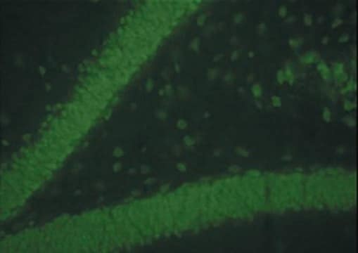
Indirect immunofluorescence of formalin fixed paraffin-embedded sections of human intestine sections: 1:10-1:20.
Immunoblotting
Optimal working dilutions must be determined by the end user.
Immunoblotting
Optimal working dilutions must be determined by the end user.
Research Category
Cell Structure
Cell Structure
Research Sub Category
Cytoskeleton
Cytoskeleton
Linkage
Replaces: 04-585
Physical form
Rabbit antiserum. Liquid containing 0.1% sodium azide as a preservative.
Storage and Stability
Maintain at -20°C in undiluted aliquots for up to 12 months. Avoid repeated freeze/thaw cycles.
Legal Information
CHEMICON is a registered trademark of Merck KGaA, Darmstadt, Germany
Disclaimer
Unless otherwise stated in our catalog or other company documentation accompanying the product(s), our products are intended for research use only and are not to be used for any other purpose, which includes but is not limited to, unauthorized commercial uses, in vitro diagnostic uses, ex vivo or in vivo therapeutic uses or any type of consumption or application to humans or animals.
Not finding the right product?
Try our Product Selector Tool.
recommended
Product No.
Description
Pricing
Storage Class Code
10 - Combustible liquids
WGK
WGK 1
Certificates of Analysis (COA)
Search for Certificates of Analysis (COA) by entering the products Lot/Batch Number. Lot and Batch Numbers can be found on a product’s label following the words ‘Lot’ or ‘Batch’.
Already Own This Product?
Find documentation for the products that you have recently purchased in the Document Library.
Yee Kit Tai et al.
Cells, 13(5) (2024-03-13)
Briefly (10 min) exposing C2C12 myotubes to low amplitude (1.5 mT) pulsed electromagnetic fields (PEMFs) generated a conditioned media (pCM) that was capable of mitigating breast cancer cell growth, migration, and invasiveness in vitro, whereas the conditioned media harvested from
Federica Maione et al.
Methods in molecular biology (Clifton, N.J.), 1267, 349-365 (2015-02-01)
Angiogenesis, the formation of neo-vessels, is one of the most important hallmarks of cancer. Tumor vasculature presents structural abnormalities such as dilatation of vessel diameter and hyper-branched and twisted pattern. A promising strategy in anticancer therapy to overcome the resistance
Richard L Benton et al.
The Journal of comparative neurology, 507(1), 1031-1052 (2007-12-20)
After traumatic spinal cord injury (SCI), disruption and plasticity of the microvasculature within injured spinal tissue contribute to the pathological cascades associated with the evolution of both primary and secondary injury. Conversely, preserved vascular function most likely results in tissue
Witold W Kilarski et al.
Laboratory investigation; a journal of technical methods and pathology, 85(5), 643-654 (2005-02-22)
Migration, proliferation and invasive growth of myofibroblasts are key cellular events during formation of granulation tissue in situations of wound healing, arteriosclerosis and tumor growth. To study the invasive phenotype of myofibroblasts, we established an assay where arterial tissue from
G P Sui et al.
BJU international, 90(1), 118-129 (2002-06-26)
To determine whether suburothelial interstitial cells of the human bladder express gap junctions, and if so, to establish their extent and composition, using immunocytochemistry, confocal microscopy and electron microscopy. Bladder tissue was obtained at cystectomy; the tissue was: (i) frozen
Our team of scientists has experience in all areas of research including Life Science, Material Science, Chemical Synthesis, Chromatography, Analytical and many others.
Contact Technical Service


