Skip to Content
MilliporeSigma
All Photos(2)

Documents

SAB4501880

Sigma-Aldrich

Anti-KI67 antibody produced in rabbit

affinity isolated antibody

Synonym(s):

Antigen KI-67, MKI67

Sign Into View Organizational & Contract Pricing


About This Item

UNSPSC Code:
12352203
NACRES:
NA.41

biological source

rabbit

conjugate

unconjugated

antibody form

affinity isolated antibody

antibody product type

primary antibodies

clone

polyclonal

form

buffered aqueous solution

mol wt

antigen 358 kDa

species reactivity

human

concentration

~1 mg/mL

technique(s)

ELISA: 1:10000
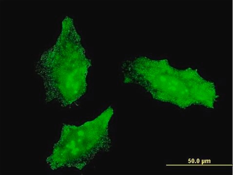
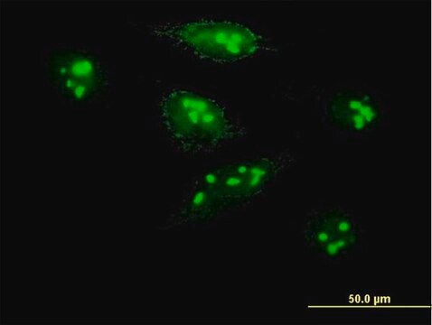
immunofluorescence: 1:100-1:500
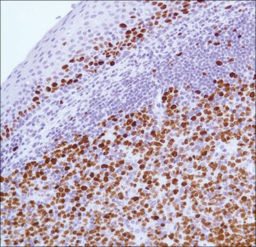
immunohistochemistry: 1:50-1:100

NCBI accession no.

UniProt accession no.

shipped in

wet ice

storage temp.

−20°C

target post-translational modification

unmodified

Gene Information

human ... MKI67(4288)

General description

Marker of proliferation Ki-67 (MKI67) is encoded by the gene mapped to human chromosome 10. The encoded protein is characterized by the phosphopeptide-binding forkhead-associated (FHA) domain and a protein phosphatase 1 (PP1)-binding site at N- terminal, 16 tandem repeats at central region and leucine and arginine (LR) pairs at C- terminal end.

Immunogen

The antiserum was produced against synthesized peptide derived from human Ki67.

Immunogen Range: 3207-3256

Application

Anti-KI67, C-Terminal antibody produced in rabbit has been used in following studies:
  • immunohistochemistry.
  • immunofluorescence staining.
  • western blotting

Biochem/physiol Actions

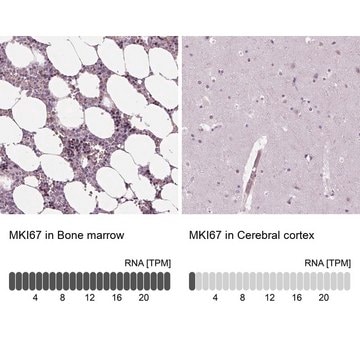
Marker of proliferation Ki-67 (MKI67), which is a part of the mitotic chromosome periphery, function as a biological surfactant to maintain individual mitotic chromosomes dispersed in the cytoplasm after nuclear envelope disassembly. Ki-67 is a vital prognostic marker in breast and prostate cancer. Expression of the encoded protein is associated with cell-proliferation rate. Ki-67 is expressed at all active phases of the cell cycle (G (1), S, G (2), and mitosis), except resting cells (G (0)). Therefore, this protein is considered as a potential cell proliferation marker.

Features and Benefits

Evaluate our antibodies with complete peace of mind. If the antibody does not perform in your application, we will issue a full credit or replacement antibody. Learn more.

Physical form

Rabbit IgG in phosphate buffered saline (without Mg2+ and Ca2+), pH 7.4, 150mM NaCl, 0.02% sodium azide and 50% glycerol.

Disclaimer

Unless otherwise stated in our catalog or other company documentation accompanying the product(s), our products are intended for research use only and are not to be used for any other purpose, which includes but is not limited to, unauthorized commercial uses, in vitro diagnostic uses, ex vivo or in vivo therapeutic uses or any type of consumption or application to humans or animals.

Not finding the right product?  

Try our Product Selector Tool.

Storage Class

12 - Non Combustible Liquids

wgk_germany

nwg

flash_point_f

Not applicable

flash_point_c

Not applicable


Certificates of Analysis (COA)

Search for Certificates of Analysis (COA) by entering the products Lot/Batch Number. Lot and Batch Numbers can be found on a product’s label following the words ‘Lot’ or ‘Batch’.

Already Own This Product?

Find documentation for the products that you have recently purchased in the Document Library.

Visit the Document Library

L Bubendorf et al.
The Journal of pathology, 178(4), 437-441 (1996-04-01)
The clinical course of prostate cancer is highly variable and cannot satisfactorily be predicted by histological criteria alone. Both tumour cell proliferation and neuroendocrine differentiation have been suggested as additional prognostic parameters, neuroendocrine differentiation being considered to enhance tumour cell
Di Wu et al.
Medical science monitor : international medical journal of experimental and clinical research, 25, 6946-6954 (2019-12-21)
BACKGROUND High blood glucose levels in diabetes result in retinal angiogenesis, which is the key feature of diabetic retinopathy. This study aimed to investigate the effects of the CXCR4 antagonist, AMD3465, on human retinal vascular endothelial cells (hRVECs) [i]in vitro[/i].
E C Inwald et al.
Breast cancer research and treatment, 139(2), 539-552 (2013-05-16)
The proliferation marker Ki-67 is one of the most controversially discussed parameters for treatment decisions in breast cancer patients. The purpose of this study was to evaluate the routine use and value of Ki-67 as a prognostic marker, and to
Sara Cuylen et al.
Nature, 535(7611), 308-312 (2016-07-01)
Eukaryotic genomes are partitioned into chromosomes that form compact and spatially well-separated mechanical bodies during mitosis. This enables chromosomes to move independently of each other for segregation of precisely one copy of the genome to each of the nascent daughter
Omar Haque et al.
Frontiers in surgery, 8, 644859-644859 (2021-07-06)
Background: A major limitation in expanding the use of donation after circulatory death (DCD) livers in transplantation is the increased risk of graft failure secondary to ischemic cholangiopathy. Warm ischemia causes thrombosis and injury to the peribiliary vascular plexus (PVP)

Our team of scientists has experience in all areas of research including Life Science, Material Science, Chemical Synthesis, Chromatography, Analytical and many others.

Contact Technical Service