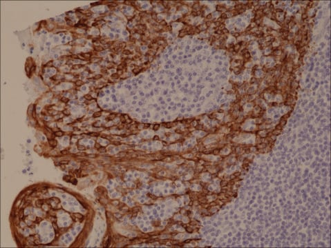
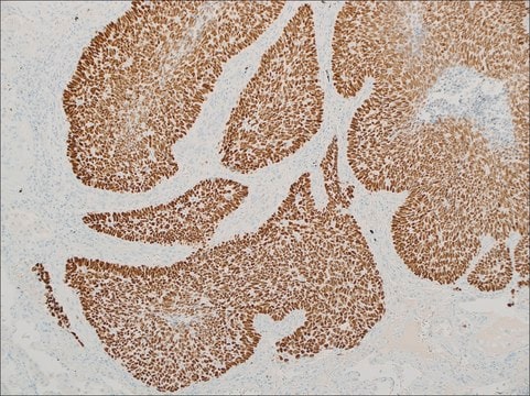
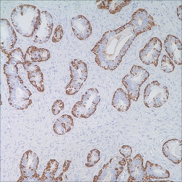
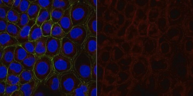
356R-1
Cytokeratin 5 & 6 (EP24 & EP67) Rabbit Monoclonal Primary Antibody
Synonym(s):
CK 5/6, cytokeratin 5/6
About This Item
Recommended Products
biological source
rabbit
Quality Level
100
500
conjugate
unconjugated
antibody form
culture supernatant
antibody product type
primary antibodies
clone
EP24 & EP67, monoclonal
description
For In Vitro Diagnostic Use in Select Regions (See Chart)
form
buffered aqueous solution
species reactivity
human
packaging
vial of 0.1 mL concentrate (356R-14)
vial of 0.1 mL concentrate Research Use Only (356R-14-RUO)
vial of 0.5 mL concentrate (356R-15)
vial of 1.0 mL concentrate (356R-16)
vial of 1.0 mL concentrate Research Use Only (356R-16-RUO)
vial of 1.0 mL pre-dilute Research Use Only (356R-17-RUO)
vial of 1.0 mL pre-dilute ready-to-use (356R-17)
vial of 7.0 mL pre-dilute ready-to-use (356R-18)
vial of 7.0 mL pre-dilute ready-to-use Research Use Only (356R-18-RUO)
manufacturer/tradename
Cell Marque™
technique(s)
immunohistochemistry (formalin-fixed, paraffin-embedded sections): 1:25-1:100 (concentrated)
control
mesothelioma, prostate, squamous cell carcinoma ((lung))
shipped in
wet ice
storage temp.
2-8°C
visualization
cytoplasmic
Gene Information
human ... KRT5(3852)
Related Categories
General description
Quality
Canada - IVD
European Union - IVD
Japan - RUO
Linkage
Physical form
Preparation Note
Note: This requires a keycode which can be found on your packaging or product label.
Download the latest released IFU
Note: This IFU may not apply to your specific product lot.
Other Notes
Legal Information
Not finding the right product?
Try our Product Selector Tool.
Storage Class
12 - Non Combustible Liquids
wgk_germany
WGK 2
flash_point_f
Not applicable
flash_point_c
Not applicable
Certificates of Analysis (COA)
Search for Certificates of Analysis (COA) by entering the products Lot/Batch Number. Lot and Batch Numbers can be found on a product’s label following the words ‘Lot’ or ‘Batch’.
Already Own This Product?
Find documentation for the products that you have recently purchased in the Document Library.
Related Content
Trusted IVDR-ready supplier of high-quality Cell Marque™ antibodies for diagnostic pathology and IHC, ensuring reliability and expert support.
Our team of scientists has experience in all areas of research including Life Science, Material Science, Chemical Synthesis, Chromatography, Analytical and many others.
Contact Technical Service


