MAB1217-I
Anti-Fmc-7 (B-Cell Lymphocyte Marker), clone Fmc-7 Antibody
clone Fmc-7, from mouse
Sign Into View Organizational & Contract Pricing
All Photos(2)
About This Item
UNSPSC Code:
12352203
eCl@ss:
32160702
NACRES:
NA.41
Recommended Products
biological source
mouse
Quality Level
antibody form
purified immunoglobulin
antibody product type
primary antibodies
clone
Fmc-7, monoclonal
species reactivity
human
technique(s)
flow cytometry: suitable
western blot: suitable
isotype
IgMκ
shipped in
wet ice
target post-translational modification
unmodified
Gene Information
human ... MS4A1(931)
Related Categories
General description
B cells are lymphocytes that play a large role in the humoral immune response as opposed to the cell-mediated immune response that is governed by T cells. The principal function of B cells is to make antibodies against soluble antigens. B cells are an essential component of the adaptive immune system. FMC-7 B-Cell Lymphocyte Marker is a 105 kDa B cell restricted antigen which is expressed on about 50% of adult human peripheral blood B cells. Upon in vivo B cell activation FMC7 expression initially increases. B cells involved in antibody secretion have lost the FMC7 determinant. The monoclonal antibody FMC7 delineates a subpopulation of B lymphocytes in normal blood. Expression of the antigen recognized by FMC7 appears to be maturation-linked, and it serves to distinguish different types of B cell leukemia.
Immunogen
HRIK [human B-lymphoblastoid cell] corresponding to human Fmc-7 (B-Cell Lymphocyte Marker).
Application
Flow Cytometry Analysis: 4.0 µg of this antibody detected Fmc-7 (B-Cell Lymphocyte Marker) in peripheral blood mononuclear cells.
This Anti-Fmc-7 (B-Cell Lymphocyte Marker) antibody is validated for use in WB, FC for the detection of Fmc-7 (B-Cell Lymphocyte Marker).
Quality
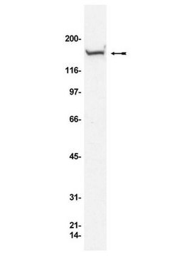
Evaluated by Western Blotting in THP-1 cell lysate.
Western Blotting Analysis: 2.0 µg/mL of this antibody detected Fmc-7 (B-Cell Lymphocyte Marker) in 10 µg of THP-1 cell lysate.
Western Blotting Analysis: 2.0 µg/mL of this antibody detected Fmc-7 (B-Cell Lymphocyte Marker) in 10 µg of THP-1 cell lysate.
Target description
~105 kDa observed. Uncharacterized band(s) may appear in some lysates.
Physical form
Format: Purified
Purified mouse monoclonal IgMκ in buffer containing 0.1M Tris, 10mM Glycine, 0.5M NaCl, and 0.1% Sodium Azide.
Other Notes
Concentration: Please refer to lot specific datasheet.
Not finding the right product?
Try our Product Selector Tool.
Storage Class
12 - Non Combustible Liquids
wgk_germany
WGK 1
flash_point_f
Not applicable
flash_point_c
Not applicable
Certificates of Analysis (COA)
Search for Certificates of Analysis (COA) by entering the products Lot/Batch Number. Lot and Batch Numbers can be found on a product’s label following the words ‘Lot’ or ‘Batch’.
Already Own This Product?
Find documentation for the products that you have recently purchased in the Document Library.
Our team of scientists has experience in all areas of research including Life Science, Material Science, Chemical Synthesis, Chromatography, Analytical and many others.
Contact Technical Service


