
DUO82064
Duolink® In Situ Microplate Nuclear Stain, Anti-Fade
Synonym(s):
in situ Proximity Ligation Assay reagent, Protein Protein Interaction Assay reagent
Sign Into View Organizational & Contract Pricing
All Photos(1)
About This Item
UNSPSC Code:
12352200
NACRES:
NA.32
Recommended Products


product line
Duolink®
Quality Level
technique(s)
proximity ligation assay: suitable
fluorescence
λex 360 nm; λem 460 nm
suitability
suitable for fluorescence-detection automated sequencing
suitable for microtiter plates
shipped in
dry ice
storage temp.
−20°C
Application
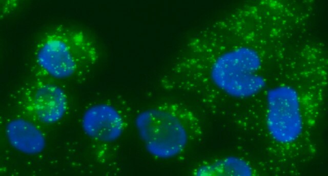
Duolink®proximity ligation assay(PLA®) allows for endogenous detection of protein interactions, post translational modifications, and protein expression levels at the single molecule level in fixed cells and tissue samples.
Use the Multiwell Plates modifications to the Duolink® In Situ Fluorescence Protocol to run an experiment with this product. A set of short instructions can also be used.
Visit our Duolink® PLA Resource Center for information on how to run a Duolink® experiment, applications, troubleshooting, and more.
To perform a complete Duolink® PLA in situ experiment you will need two primary antibodies (PLA, IHC, ICC or IF validated) that recognize two target epitopes. Other necessary reagents include a pair of PLA probes from different species (one PLUS and one MINUS), detection reagents, wash buffers, and mounting medium. Note that the primary antibodies must come from the same species as the Duolink® PLA probes. Analysis is carried out using standard immunofluorescence assay equipment.
Specificity
Duolink® In Situ Microplate Nuclear Stain and Anti-Fade are intended to be used after staining cells with Duolink® In Situ in microtiter plates. See the datasheet for more information.
Application Note
Two primary antibodies raised in different species are needed. Test your primary antibodies (IgG-class, mono- or polyclonal) in a standard immunofluorescence (IF), immunohistochemistry (IHC) or immunocytochemistry (ICC) assay to determine the optimal fixation, blocking, and titer conditions. Duolink® in situ reagents are suitable for use on fixed cells, cytospin cells, cells grown on slide, formalin-fixed, paraffin embedded (FFPE), or tissue (fresh or frozen). No minimum number of cells is required.
Let us do the work for you, learn more about our Custom Service Program to accelerate your Duolink® projects
View full Duolink® product list
Features and Benefits
- No overexpression or genetic manipulation required
- High specificity (fewer false positives)
- Single molecule sensitivity due to rolling circle amplification
- Relative quantification possible
- No special equipment needed
- Quicker and simpler than FRET
- Increased accuracy compared to co-IP
- Publication-ready results
Legal Information
Duolink is a registered trademark of Merck KGaA, Darmstadt, Germany
PLA is a registered trademark of Merck KGaA, Darmstadt, Germany

Signal Word
Warning
Hazard Statements
Precautionary Statements
Hazard Classifications
Aquatic Chronic 3 - Skin Sens. 1
Storage Class Code
12 - Non Combustible Liquids
Flash Point(F)
Not applicable
Flash Point(C)
Not applicable
Choose from one of the most recent versions:
Already Own This Product?
Find documentation for the products that you have recently purchased in the Document Library.
Customers Also Viewed


Jeffrey J Raizer et al.
Cancer, 116(22), 5297-5305 (2010-07-29)
The authors evaluated a 3-week schedule of bevacizumab in patients with recurrent high-grade glioma (HGG). Patients received bevacizumab 15 mg/kg every 3 weeks and were evaluated every 6 weeks until tumor progression. Tissue correlates were used to quantify tumor content
Jaclyn J Renfrow et al.
Neuro-oncology, 13(8), 880-885 (2011-07-30)
We present a novel methodology combining traditional fluorescent in situ hybridization with an in situ protein detection technology called proximity ligation assay. This method has potential to perform a detailed analysis of the relationship between gene status and corresponding protein
Thomas W Bonagura et al.
Endocrinology, 153(6), 2897-2906 (2012-04-13)
We previously showed that advancing the increase in estradiol levels from the second to the first third of baboon pregnancy suppressed placental extravillous trophoblast (EVT) invasion and remodeling of the uterine spiral arteries. Cell culture studies show that vascular endothelial
Ajay K Yadav et al.
JAMA, 302(3), 276-289 (2009-07-16)
Glioblastomas--uniformly fatal brain tumors--often have both monosomy of chromosome 10 and gains of the epidermal growth factor receptor (EGFR) gene locus on chromosome 7, an association for which the mechanism is poorly understood. To assess whether coselection of EGFR gains
Charles Lu et al.
PloS one, 7(4), e34833-e34833 (2012-05-05)
Tumor suppressor gene TUSC2/FUS1 (TUSC2) is frequently inactivated early in lung cancer development. TUSC2 mediates apoptosis in cancer cells but not normal cells by upregulation of the intrinsic apoptotic pathway. No drug strategies currently exist targeting loss-of-function genetic abnormalities. We
Our team of scientists has experience in all areas of research including Life Science, Material Science, Chemical Synthesis, Chromatography, Analytical and many others.
Contact Technical Service