Skip to Content
Merck
All Photos(2)

Key Documents

C7617

Sigma-Aldrich

Anti-Calnexin antibody, Mouse monoclonal

enhanced validation

clone TO-5, purified from hybridoma cell culture

Synonym(s):

Anti-CANX, Anti-CNX

Sign Into View Organizational & Contract Pricing


About This Item

MDL number:
UNSPSC Code:
12352203
NACRES:
NA.44

biological source

mouse

Quality Level

conjugate

unconjugated

antibody form

purified immunoglobulin

antibody product type

primary antibodies

clone

TO-5, monoclonal

form

buffered aqueous solution

mol wt

antigen ~90 kDa

species reactivity

human

enhanced validation

independent
Learn more about Antibody Enhanced Validation

concentration

~2 mg/mL

technique(s)

flow cytometry: suitable
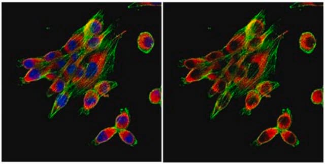
immunocytochemistry: suitable
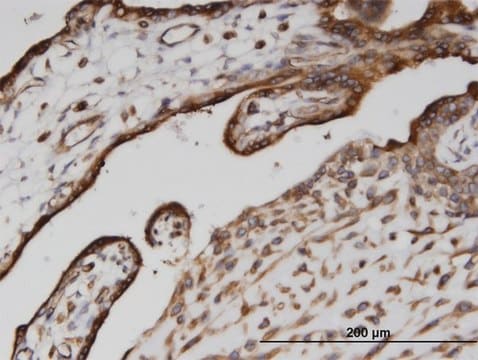
immunohistochemistry: suitable
indirect ELISA: suitable
western blot: 0.5-1.0 μg/mL using total cell extract of HeLa cells.

isotype

IgG1

UniProt accession no.

shipped in

dry ice

storage temp.

−20°C

target post-translational modification

unmodified

Gene Information

human ... CANX(821)

General description

Anti-Calnexin antibody, Mouse monoclonal (mouse IgG1 isotype) is derived from the hybridoma TO-5 produced by the fusion of mouse myeloma cells and splenocytes from BALB/c mice. Calnexin contains a long (461 amino acids) N-terminal Ca2+-binding domain extending into the lumen of the endoplasmic reticulum (ER), a short (22 amino acids) trans-membrane segment and an acidic cytosolic domain (96 amino acids).
Calnexin (p88, IP90) is a calcium-binding, type I integral membrane protein, localized primarily in the endoplasmic reticulum (ER). Calnexin binds newly synthesized glycoproteins and misfolded proteins and is believed to play a critical role in quality control processes during protein synthesis and folding. Calnexin acts as a lectin like chaperone that binds oligosaccharide residues of newly synthesized N-linked glycoproteins. The lectin specificity of calnexin and its soluble homologue calreticulin has been identified as high mannose oligosaccharides terminating in monoglucosyl residues linked through α1-3. Calnexin has been shown to be associated with several cell surface proteins, including MHC class I heavy chain, T-cell receptor (TCR), and B cell membrane immunoglobulin during translocation through the ER. It also forms complexes with other resident ER proteins involved in Ca2+-dependent retention of proteins.

Specificity

Mouse monoclonal clone TO-5 anti-Calnexin antibody recognizes human Calnexin, ~90 kDa.

Application

Anti-Calnexin antibody, Mouse monoclonal has been used in:
  • enzyme-linked immunosorbent assay (ELISA)
  • immunoblotting
  • flow cytometry
  • immunocytochemistry
  • immunohistochemistry
  • immunofluorescence analysis

Physical form

Solution in 0.01 M phosphate buffered saline, pH 7.4, containing 15 mM sodium azide.

Disclaimer

Unless otherwise stated in our catalog or other company documentation accompanying the product(s), our products are intended for research use only and are not to be used for any other purpose, which includes but is not limited to, unauthorized commercial uses, in vitro diagnostic uses, ex vivo or in vivo therapeutic uses or any type of consumption or application to humans or animals.

Not finding the right product?  

Try our Product Selector Tool.

Storage Class Code

10 - Combustible liquids

WGK

WGK 3

Flash Point(F)

Not applicable

Flash Point(C)

Not applicable


Certificates of Analysis (COA)

Search for Certificates of Analysis (COA) by entering the products Lot/Batch Number. Lot and Batch Numbers can be found on a product’s label following the words ‘Lot’ or ‘Batch’.

Already Own This Product?

Find documentation for the products that you have recently purchased in the Document Library.

Visit the Document Library

E Giacomini et al.
Human reproduction (Oxford, England), 36(8), 2249-2274 (2021-07-01)
Are uterine fluid-derived extracellular vesicles (UF-EVs) a 'liquid biopsy' reservoir of biomarkers for real-time monitoring of endometrial status? The transcriptomic cargo of UF-EVs reflects the RNA profile of the endometrial tissue as well as changes between the non-receptive and the
Identification of RUVBL1 and RUVBL2 as Novel Cellular Interactors of the Ebola Virus Nucleoprotein
Morwitzer MJ, et al.
Viruses, 11(4), 372-372 (2019)
Aleksandr Treyer et al.
Molecular biology of the cell, 27(14), 2259-2271 (2016-05-27)
For several decades, the trans-Golgi network (TGN) was considered the most distal stop and hence the ultimate protein-sorting station for distinct apical and basolateral transport carriers that reach their respective surface domains in the direct trafficking pathway. However, recent reports
Begomoviral Movement Protein Effects in Human and Plant Cells: Towards New Potential Interaction Partners
Krapp S, et al.
Viruses, 9(11), 334-334 (2017)
Induction of a massive endoplasmic reticulum and perinuclear space expansion by expression of lamin B receptor mutants and the related sterol reductases TM7SF2 and DHCR7
Zwerger M, et al.
Molecular Biology of the Cell, 21(2), 354-368 (2010)

Our team of scientists has experience in all areas of research including Life Science, Material Science, Chemical Synthesis, Chromatography, Analytical and many others.

Contact Technical Service