Skip to Content
Merck
All Photos(2)

Key Documents

A4450

Sigma-Aldrich

Monoclonal Anti-β1 and β2-Adaptins antibody produced in mouse

clone 100/1, ascites fluid

Synonym(s):

Anti-AP-1 and AP-2

Sign Into View Organizational & Contract Pricing


About This Item

MDL number:
UNSPSC Code:
12352203
NACRES:
NA.41

biological source

mouse

Quality Level

conjugate

unconjugated

antibody form

ascites fluid

antibody product type

primary antibodies

clone

100/1, monoclonal

contains

15 mM sodium azide

species reactivity

human, bovine, rat

technique(s)

indirect immunofluorescence: suitable
microarray: suitable
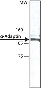
western blot: 1:200 using rat brain extract

isotype

IgG1

UniProt accession no.

shipped in

dry ice

storage temp.

−20°C

target post-translational modification

unmodified

Gene Information

General description

Monoclonal Anti- β1 and β2-Adaptins (AP-1 & AP-2)(mouse IgG1 isotype) is derived from the 100/1 hybridoma produced by the fusion of mouse myeloma cells and splenocytes from an immunized mouse. Adaptins AP-1 and AP-2 are mapped to human chromosome 22q12.2 and 17q12, respectively. AP-1 adaptor consists of four subunits. It includes two large adaptins (one each of γ/α/δ/ε and β1−4 , one medium adaptin (μ1−4), and one small adaptin (ς1−4. Similarly, the AP-2 is made up of two 100-110 kDa polypeptides, termed α and β2 (formerly β) subunits, F2 , and s2 subunit. α and β2 subunits (α and β 2).

Immunogen

AP-2 adaptor protein from bovine brain.

Application

Immunofluorescence of MDCK cells fixed in methanol was performed using anti-AP1 at a dilution of 1:250 and also western blot at a dilution of 1:1000.
Monoclonal Anti-β1 and β2-Adaptins antibody produced in mouse has been used in immunoblotting and immunohistology.
Mouse monoclonal anti-β1 and β2-adaptins can be used for western blot (1:200) assays using rat brain extract. The antibody is also suitable for use in indirect immunofluorescence and microarray analyses. Furthermore, monoclonal anti-β1/β2-adaptins have been used for double staining methods to detect clathrin-coated regions of the plasma membrane in rat brain cells.

Biochem/physiol Actions

β1and β2 adaptins are highly homologous which suggests that the β -type adaptor chains may mediate the interaction of their adaptors with the highly conserved clathrin molecule. Monoclonal antibodies reacting specifically against adaptor proteins are useful tools for studies on the intracellular distribution and structural relationship of adaptor complexes.
Clathrin-coated vesicles facilitate the selective internalization of membrane receptors for lysosomal enzymes from the trans-Golgi network to a pre-lysosomal compartment. Besides clathrin, coated vesicle populations contain the adaptor complexes AP-1 and AP-2, also known as HA-I (HA1) adaptor and HA-II (HA2) adaptor or Assemble Protein 1 (AP1) and Assemble Protein 2 (AP2), respectively. These adaptor proteins mediate the interaction between clathrin and membrane components.

Physical form

The antibody is provided as ascites fluid with 0.1% sodium azide as a preservative.

Disclaimer

Unless otherwise stated in our catalog or other company documentation accompanying the product(s), our products are intended for research use only and are not to be used for any other purpose, which includes but is not limited to, unauthorized commercial uses, in vitro diagnostic uses, ex vivo or in vivo therapeutic uses or any type of consumption or application to humans or animals.

Not finding the right product?  

Try our Product Selector Tool.

Storage Class Code

10 - Combustible liquids

WGK

WGK 3

Flash Point(F)

Not applicable

Flash Point(C)

Not applicable


Choose from one of the most recent versions:

Certificates of Analysis (COA)

Lot/Batch Number

Don't see the Right Version?

If you require a particular version, you can look up a specific certificate by the Lot or Batch number.

Already Own This Product?

Find documentation for the products that you have recently purchased in the Document Library.

Visit the Document Library

Asja Nürnberger et al.
The journal of histochemistry and cytochemistry : official journal of the Histochemistry Society, 52(12), 1647-1655 (2004-11-24)
The dopamine D3 receptor (D3R), intensively studied in neuroscience, also plays an important role in the regulation of renal and cardiovascular function. In contrast to functional findings, less information is available on its localization in the kidney. Neither RT-PCR studies
Adaptins: the final recount
Boehm M and Bonifacino JS
Molecular Biology of the Cell, 12(10), 2907-2920 (2001)
Otitis media: a genome-wide linkage scan with evidence of susceptibility loci within the 17q12 and 10q22. 3 regions
Casselbrant ML, et al.
BMC Medical Genetics, 10(1), 85-85 (2009)
The structure and function of the beta2-adaptin appendage domain
Owen DJ, et al.
Molecular Biology of the Cell, 19(16), 4216-4227 (2000)
Molecular and genetic profiles of radiographically defined de novo meningiomas
Kitamura Y, et al.
Journal of Neuro-Oncology, 108, 37-44 (2012)

Our team of scientists has experience in all areas of research including Life Science, Material Science, Chemical Synthesis, Chromatography, Analytical and many others.

Contact Technical Service