


Following citation support the suitability of the antibody for flow cytometry " Microvasc Res. 2004 Mar;67(2):168-81".
F4771
Monoclonal Anti-Fibroblast Surface Protein antibody produced in mouse
clone 1B10, ascites fluid
Synonym(s):
Fibroblast Surface Protein Antibody, Fibroblast Surface Protein Antibody - Monoclonal Anti-Fibroblast Surface Protein antibody produced in mouse
Sign In to View Organizational & Contract Pricing.
Select a Size
Change View
| Size/SKU | Availability | Price |
|---|---|---|
0.2 mL | Please contact Customer Service for Availability | PLN 1,860.00 |
About This Item
Conjugate:
unconjugated
Clone:
1B10, monoclonal
Application:
FACS, IHC (f), complement-mediated cytotoxicity assay
Citations:
25
PLN 1,860.00
Please contact Customer Service for Availability
A recombinant, preservative-free antibody is available for your target. Try ZMS1048
biological source
mouse
Quality Segment
conjugate
unconjugated
antibody form
ascites fluid
antibody product type
primary antibodies
clone
1B10, monoclonal
mol wt
antigen 43-72-80 kDa
contains
15 mM sodium azide
species reactivity
human
technique(s)
complement-mediated cytotoxicity assay: 1:500, flow cytometry: suitable, immunohistochemistry (frozen sections): suitable
isotype
IgM
shipped in
dry ice
storage temp.
−20°C
target post-translational modification
unmodified
General description
Monoclonal Anti-Human Fibroblast Surface Protein (mouse IgM isotype) is derived from the 1B10 hybridoma1 produced by the fusion of mouse myeloma cells and splenocytes from BALB/c mice immunized with cultured human thymic fibroblasts. Human fibroblast surface protein is a fibroblast antigen that is expressed mainly on human synovial, foreskin and thymic fibroblasts and is absent on human epithelial cells and lymphocytes.
This fibroblast antigen has been demonstrated on human synovial, mammary, foreskin and thymic fibroblasts and in malignant fibrosarcoma tissue. The antigen is both cell membrane and lysosome associated.
Immunogen
cultured human thymic fibroblasts
Application
Applications in which this antibody has been used successfully, and the associated peer-reviewed papers, are given below.
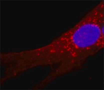
Immunofluorescence (1 paper)
Immunohistochemistry (1 paper)
Immunofluorescence (1 paper)
Immunohistochemistry (1 paper)
Monoclonal Anti-Fibroblast Surface Protein antibody produced in mouse has been used in:
- immunofluorescence
- flow cytometry
- immunohistochemistry
- immunoblotting
Monoclonal anti-fibroblast surface protein antibody can be used in flow cytometry of human fibroblasts and monocytes. It is also useful in immunoblotting and western blotting.
Biochem/physiol Actions
Monoclonal Anti-Fibroblast Surface Protein antibody is also used to remove fibroblasts from human cultured cells by inhibiting fibroblasts adherence to the culture vessel or by cytotoxic effect in the presence of rabbit complement. In addition, apart from the functional significance of cells characteristics, the characterization of cell lines is important for correlation of cultures with tissue origin, identification of the lineage to which the cells belong or the precursor status of the cells. Monoclonal antibodies to fibroblast surface protein should provide a powerful tool for control of fibroblast growth in a variety of human cell culture systems, such as thymic epithelial (TE) cell cultures allowing the growth of highly enriched populations of the TE cells.
Analysis Note
Working dilution is at least 1:500 by complement-mediated microcytotoxicity assay.
Disclaimer
Unless otherwise stated in our catalog or other company documentation accompanying the product(s), our products are intended for research use only and are not to be used for any other purpose, which includes but is not limited to, unauthorized commercial uses, in vitro diagnostic uses, ex vivo or in vivo therapeutic uses or any type of consumption or application to humans or animals.
1 of 1
This Item | |||
|---|---|---|---|
| antibody form ascites fluid | antibody form purified immunoglobulin | antibody form ascites fluid | antibody form ascites fluid |
| clone 1B10, monoclonal | clone FB-8, monoclonal | clone COL-1, monoclonal | clone SVP-38, monoclonal |
| conjugate unconjugated | conjugate unconjugated | conjugate unconjugated | conjugate unconjugated |
| biological source mouse | biological source mouse | biological source mouse | biological source mouse |
| species reactivity human | species reactivity bovine, human | species reactivity bovine, human, pig, rat, rabbit, deer | species reactivity pig, rat, human, guinea pig |
| technique(s) complement-mediated cytotoxicity assay: 1:500, immunohistochemistry (frozen sections): suitable, flow cytometry: suitable | technique(s) dot blot: suitable, indirect ELISA: suitable, neutralization: 5 μg/mL using 1 unit recombinant human FGF2 in mouse 3T3 cells, radioimmunoassay: suitable, western blot: suitable | technique(s) dot blot: suitable, immunohistochemistry (frozen sections): 1:2000 using human or other mammalian frozen sections, indirect ELISA: suitable | technique(s) immunohistochemistry (formalin-fixed, paraffin-embedded sections): suitable, immunohistochemistry (frozen sections): 1:200 using rat cerebellum, indirect ELISA: suitable, western blot: suitable |
Still not finding the right product?
Try our Product Selector Tool to narrow your options
Storage Class
10 - Combustible liquids
wgk
nwg
flash_point_f
Not applicable
flash_point_c
Not applicable
Choose from one of the most recent versions:
Already Own This Product?
Find documentation for the products that you have recently purchased in the Document Library.
-
Is there specific data or a scientific paper available to support the claim that this antibody is suitable for flow cytometry?
1 answer-
Helpful?
-