Skip to Content
Merck
All Photos(1)

Documents

MABN15

Sigma-Aldrich

Anti-Rhodopsin Antibody, clone 4D2

clone 4D2, from mouse

Synonym(s):

rhodopsin, rhodopsin (opsin 2, rod pigment) (retinitis pigmentosa 4, autosomal dominant), retinitis pigmentosa 4, autosomal dominant, opsin 2, rod pigment

Sign Into View Organizational & Contract Pricing


About This Item

UNSPSC Code:
12352203
eCl@ss:
32160702
NACRES:
NA.41

biological source

mouse

Quality Level

antibody form

purified immunoglobulin

antibody product type

primary antibodies

clone

4D2, monoclonal

species reactivity

rat, human, fish

species reactivity (predicted by homology)

mouse (based on 100% sequence homology)

technique(s)

ELISA: suitable
immunocytochemistry: suitable
immunohistochemistry: suitable
western blot: suitable

isotype

IgG2bκ

NCBI accession no.

UniProt accession no.

shipped in

wet ice

target post-translational modification

unmodified

Gene Information

human ... RHO(6010)
rat ... Rho(24717)

General description

Rhodopsin is a multi-pass, G-protein-coupled membrane receptor protein belonging to the G-protein coupled receptor 1 family. Found within rod photoreceptor cells, rhodopsin is activated by light photons in order for the visual transduction cascade to initialize. Rhodopsin is necessary to the forming of images in low light and for the viability of photoreceptor cells after birth. Mutations in rhodopsin expression are the most widely known cause of the human disease retinitis pigmentosa (RP) resulting in gradual loss of vision. At present, there are over 100 known rhodopsin mutations.

Specificity

This antibody recognizes Rhodopsin at the N-terminus.

Immunogen

Epitope: N-terminus
Rat rod outer segments corresponding to rat Rhodopsin at the N-terminus.

Application

Anti-Rhodopsin Antibody, clone 4D2 is an antibody against Rhodopsin for use in IH, WB, IC, ELISA.
Research Category
Neuroscience
Research Sub Category
Sensory & PNS
Western Blot Analysis: A previous lot of this antibody detected Rhodopsin by WB in goldfish retina lysate, as reported by Knight K, et al.

Immunocytochemistry: A previous lot of this antibody detected Rhodopsin by ICC in goldfish retina tissue, as reported by Knight K, et al

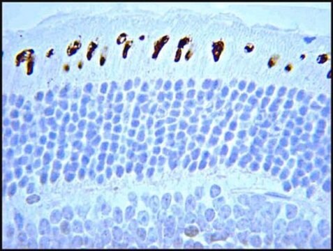
Immunohistochemistry Analysis: 1:500 dilution of this antibody has been shown to detect Rhodopsin in human eye tissue.

ELISA: Specificity of this antibody, from a different lot, was validated in a competitive ELISA, as reported by Laird D, et al.

Quality

Evaluated by Immunohistochemistry in rat eye tissue.

Immunohistochemistry Analysis: 1:500 dilution of this antibody detected Rhodopsin in rat eye tissue.

Target description

Expected at ~ 39 kDa

Physical form

Format: Purified
Protein G Purified
Purified mouse monoclonal IgG2bκ in buffer containing 0.1 M Tris-Glycine (pH 7.4), 150 mM NaCl with 0.05% sodium azide.

Storage and Stability

Stable for 1 year at 2-8°C from date of receipt.

Analysis Note

Control
Rat eye tissue

Other Notes

Concentration: Please refer to the Certificate of Analysis for the lot-specific concentration.

Disclaimer

Unless otherwise stated in our catalog or other company documentation accompanying the product(s), our products are intended for research use only and are not to be used for any other purpose, which includes but is not limited to, unauthorized commercial uses, in vitro diagnostic uses, ex vivo or in vivo therapeutic uses or any type of consumption or application to humans or animals.

Not finding the right product?  

Try our Product Selector Tool.

Storage Class Code

12 - Non Combustible Liquids

WGK

WGK 1

Flash Point(F)

Not applicable

Flash Point(C)

Not applicable


Certificates of Analysis (COA)

Search for Certificates of Analysis (COA) by entering the products Lot/Batch Number. Lot and Batch Numbers can be found on a product’s label following the words ‘Lot’ or ‘Batch’.

Already Own This Product?

Find documentation for the products that you have recently purchased in the Document Library.

Visit the Document Library

Zhe Zhang et al.
The Journal of biological chemistry, 295(34), 12153-12166 (2020-07-09)
Pheromones play critical roles in habitat identification and reproductive behavior synchronization in the sea lamprey (Petromyzon marinus). The bile acid 3-keto petromyzonol sulfate (3kPZS) is a major component of the sex pheromone mixture from male sea lamprey that induces specific
Kayvan Samimi et al.
Biomedical optics express, 13(6), 3476-3492 (2022-07-06)
Photoreceptors are the key functional cell types responsible for the initiation of vision in the retina. Phototransduction involves isomerization and conversion of vitamin A compounds, known as retinoids, and their recycling through the visual cycle. We demonstrate a functional readout
Liang Jia et al.
The Journal of biological chemistry, 297(5), 101268-101268 (2021-10-04)
Biogenic amines activate G-protein-coupled receptors (GPCRs) in the central nervous system in vertebrate animals. Several biogenic amines, when excreted, stimulate trace amine-associated receptors (TAARs), a group of GPCRs in the main olfactory epithelium, and elicit innate behaviors. How TAARs recognize
Alireza Badiei et al.
Experimental eye research, 181, 72-84 (2019-01-18)
Chronic exposure of the retina to light and high concentrations of polyunsaturated fatty acid in photoreceptor cells make this tissue susceptible to oxidative damage. As retinal degenerative diseases are associated with photoreceptor degeneration, the antioxidant activity of both hydrogen sulfide
Roni A Hazim et al.
Experimental eye research, 179, 18-24 (2018-10-20)
Human RPE cell lines, especially the ARPE-19 cell line, are widely-used in eye research, as well as general epithelial cell studies. In comparison with primary RPE cells, they offer relative convenience and consistency, but cultures derived from these lines are typically

Our team of scientists has experience in all areas of research including Life Science, Material Science, Chemical Synthesis, Chromatography, Analytical and many others.

Contact Technical Service