immunocytochemistry: 1:200 using methanol-fixed, dog MDCK and human MCF7 cellc microarray: suitable western blot: 1:1,000 using whole cell extract of human epidermal carcinoma A431 cell line and cytosolic fraction of rat embryonic brain.
α-E-Catenin is a predominant subtype of α-catenin. It is widely expressed but at low levels in the nervous system. Alternative spliced variants of α-E-Catenin include α1- and α2-E-catenin.
The catenins (α, β, γ) are cytoplasmic proteins found in varying abundance in many developing and adult tissues.
Immunogen
synthetic peptide corresponding to a region located near the C-terminus of human α-E-catenin (amino acids 873-887). This sequence is identical in mouse and Xenopus α-E-catenin. It is not found in α-N-catenin, β-catenin, and γ-catenin.
Application
Anti-α-E-Catenin antibody produced in rabbit has been used in western blotting and immunocytochemistry.
Biochem/physiol Actions
Catenins bind directly or indirectly to the conserved cytoplasmic tail domain of the cell adhesion adherins. The association of catenins to cadherins produces a complex, which is linked to the actin filament network. Catenins/cadherin complexes play an important role in mediating cell adhesion, transduction of cell-cell contact positional signals to the cell interior, and may play a crucial role in cell differentiation. The linkage of the epithelial cadherin /uvomorulin to actin is essential for the cell binding function of this cadherin. α-Catenin (CAP102, 102 kDa), originally described as an E-cadherin associated protein, has been shown to associate with other members of the cadherin family members, N-cadherin and P-cadherin. Within its conserved region α-catenin shows 30% identity to vinculin.
Physical form
Solution in 0.01 M phosphate buffered saline, pH 7.4, containing 15 mM sodium azide.
Disclaimer
Unless otherwise stated in our catalog or other company documentation accompanying the product(s), our products are intended for research use only and are not to be used for any other purpose, which includes but is not limited to, unauthorized commercial uses, in vitro diagnostic uses, ex vivo or in vivo therapeutic uses or any type of consumption or application to humans or animals.
Mitotic spindle orientation (SO) is a conserved mechanism that governs cell fate and tissue morphogenesis. In the developing epidermis, a balance between self-renewing symmetric divisions and differentiative asymmetric divisions is necessary for normal development. While the cellular machinery that executes
p120 catenin is required for normal renal tubulogenesis and glomerulogenesis
The establishment of tissue architecture requires coordination between distinct processes including basement membrane assembly, cell adhesion, and polarity; however, the underlying mechanisms remain poorly understood. The actin cytoskeleton is ideally situated to orchestrate tissue morphogenesis due to its roles in
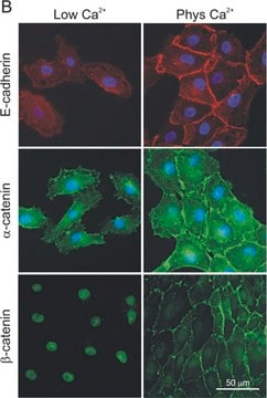
Cadherins are a family of transmembrane glycoproteins which play a key role in Ca(2+)-dependent cell-cell adhesion. Cytoplasmic domains of these molecules are anchored to the cell cytoskeleton and are required for cadherin function. To elucidate how the function of cadherins
Our team of scientists has experience in all areas of research including Life Science, Material Science, Chemical Synthesis, Chromatography, Analytical and many others.