General description
Fibronectin (FN) is an extracellular matrix protein composed of two nearly-identical disulfide-bound polypeptides with typical molecular weights of 220-280 kDa. Cellular fibronectin is structurally and antigenically similar to cold insoluble globulin from plasma and antibodies to either form usually cross-react. Careful analysis of the FN molecule indicates that it contains several functionally and structurally distinct domains which may bind to cell surfaces and to a variety of molecules such as collagen, heparin, gelatin, fibrin and DNA. FN′s play an important role in diverse biological phenomena including cell adhesion, cell migration, hemostasis and thrombosis, wound healing and the ability to induce a more normal phenotype in transformed cells.
Monoclonal Anti-Fibronectin (mouse IgG1 isotype) is derived from the hybridoma produced the fusion of mouse myeloma cells and splenocytes from immunized mice. Fibronectin (FN) belongs to glycoprotein family widely expressed in many tissues. FN is an extracellular matrix protein composed of two nearly-identical disulfide-bound polypeptides with typical molecular weights of 220-240 kDa. There are three isoforms of fibronectin, that includes cellular FN, plasma FN and fetal FN. Cellular fibronectin is structurally and antigenically similar to cold insoluble globulin from plasma and antibodies to either form usually cross-react. Careful analysis of the FN molecule indicates that it contains several functionally and structurally distinct domains which may bind to cell surfaces and to a variety of molecules such as collagen, heparin, gelatin, fibrin and DNA. FN gene in human chromosome is mapped to 2q35.
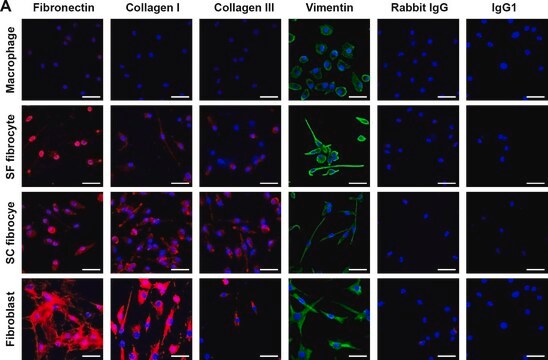
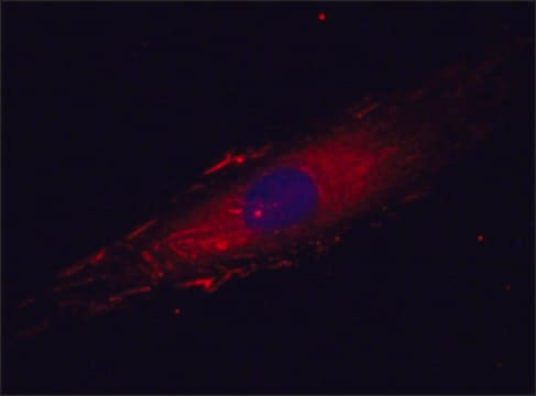
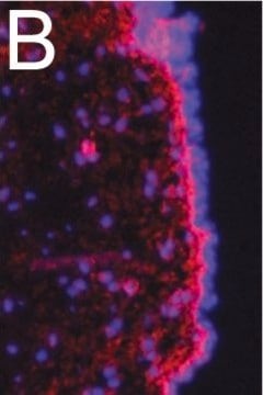
Mouse monoclonal clone FN-15 anti-Fibronectin antibody labels fibronectin in the pericellular extracellular matrix of cultured human fibroblasts using indirect immunofluorescent methods. It also labels fibronectin in chicken and mouse cells. The FN-15 antibody specifically stains fibronectin in immunoblotting, and in an ELISA is reactive with human plasma fibronectin.
Immunogen
fibronectin from human plasma
Application
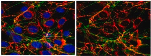
Monoclonal Anti-Fibronectin may be used to specifically localize fibronectin in human cell culture and in tissue sections by immunoblotting, immunohistochemical techniques and to stain fibronectin in hydrogels.
Mouse monoclonal clone FN-15 anti-Fibronectin antibody may be used to specifically localize fibronectin in human cell culture and in tissue sections by immunoblotting and immunohistochemical techniques. Monoclonal antibodies reacting specifically with FN may be used to localize FN in human cell cultures, in tissue sections and in plasma.
Biochem/physiol Actions
Fibronectin (FN) play an important role in diverse biological phenomena including cell adhesion, cell migration, hemostasis and thrombosis, wound healing and the ability to induce a more normal phenotype in transformed cells. FN is upregulated in many cancers. FN increases the expression of matrix metalloproteinases (MMPs), promoting cancer cell invasion and metastasis.
Disclaimer
Unless otherwise stated in our catalog or other company documentation accompanying the product(s), our products are intended for research use only and are not to be used for any other purpose, which includes but is not limited to, unauthorized commercial uses, in vitro diagnostic uses, ex vivo or in vivo therapeutic uses or any type of consumption or application to humans or animals.
