Skip to Content
Merck
All Photos(2)

Documents

MABS1152

Sigma-Aldrich

Anti-ROR2 Antibody, clone Nt 2535-2835

clone Nt2535-2835, from mouse

Synonym(s):

Tyrosine-protein kinase transmembrane receptor ROR2, mRor2, Ntrkr2, Neurotrophic tyrosine kinase receptor-related 2

Sign Into View Organizational & Contract Pricing


About This Item

UNSPSC Code:
12352203
eCl@ss:
32160702
NACRES:
NA.41

biological source

mouse

Quality Level

antibody form

purified immunoglobulin

antibody product type

primary antibodies

clone

Nt2535-2835, monoclonal

species reactivity

mouse, human

technique(s)

ELISA: suitable
immunohistochemistry (formalin-fixed, paraffin-embedded sections): suitable
western blot: suitable

isotype

IgG1κ

UniProt accession no.

shipped in

wet ice

target post-translational modification

unmodified

Gene Information

human ... ROR2(4920)

Related Categories

General description

Tyrosine-protein kinase transmembrane receptor ROR2 (EC 2.7.10.1; UniProt Q9Z138; also known as mRor2, Ntrkr2, Neurotrophic tyrosine kinase receptor-related 2) is encoded by the Ror2 gene (Gene ID 26564) in murine species. Ror2 is a receptor tyrosine kinase that binds Wnt5a and negatively regulates canonical Wnt signaling. Although in C. elegans and Xenopus Ror2 is reported to modulate Wnt signaling nonautonomously by preventing Wnt from stimulating adjacent cells, studies conducted in mammalian cells suggest that Ror2 cytoplasmic domain is crucial for its function and Ror2 inhibits Wnt signaling in a cell-autonomous manner. Ror2 and the closely related Ror1 play essential roles during development. Human ROR2 gene mutations have been associated with dominant Brachydactyly B and recessive Robinow syndrome. Ror2-null mice exhibit skeletal abnormalities similar to the human syndromes. In addition, these mice display heart and lung defects and die perinatally. Ror2 is initially translated with an N-terminal signal sequence (a.a. 1-33), the removal of which yields the mature receptor with an extracellular region (a.a. 34-403), a transmembrane segment (a.a. 404-424), followed by the cytoplasmic region (a.a. 425-944) that contains the kinase domain (a.a. 473-746), a Pro-rich domain (a.a. 784-857) sandwiched between two Ser/Thr-rich domains (a.a. 753-782 and a.a. 859-882).

Specificity

Clone Nt 2535-2835 is specific to the mouse and human ROR2, but does not detect mouse ROR1. Clone Nt 2535-2835 exclusively detects ROR2 protein in paraffin-embedded tissues (Mikels, A., et al. (2009). J. Biol. Chem. 284(44):30167-30176).

Immunogen

Epitope: Second Ser/Thr-rich domain.
Recombinant protein corresponding to the second Ser/Thr-rich domain of mouse mRor2.

Application

Anti-ROR2 Antibody, clone Nt 2535-2835 is an antibody against ROR2 for use in Western Blotting, Immunohistochemistry, Immunohistochemistry (Paraffin), ELISA.
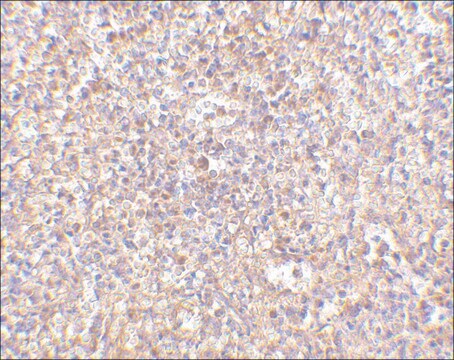
Immunohistochemistry Analysis: A representative lot detected Ror2 immunoreactivity in parafffin-embedded leiomyosarcoma (LMS) tumor, lung cancer, and breast cancer tissue sections (Courtesy of Dr. Roel Nusse, Stanford University, CA).
Immunohistochemistry Analysis: A representative lot detected Ror2 expression in various regions of wild-type and Ror2+/-, but not Ror2-/-, mouse E13.5 embryos by both fluorescent and non-fluorescent immunohistochemistry using paraformaldehyde-fixed, paraffin-embedded whole mount sections (Mikels, A., et al. (2009). J. Biol. Chem. 284(44):30167-30176).
Immunohistochemistry Analysis: A representative lot detected Ror2 expression in the dermis of wild-type, but not Ror2-knockout mouse skin from E16.5 mouse embryos using paraformaldehyde-fixed, paraffin-embedded whole mount sections (van Amerongen, R., et al. (2012). Dev. Biol. 369(1):101-114).
Western Blotting Analysis: A representative lot detected full-length murine Ror2, as well as Ror2 deletion constructs lacking the first Ser/Thr-rich domain (ST1; a.a 753-782) or the Pro-rich domain (a.a. 784-857), but not Ror2 deletion mutants lacking the ST2 (a.a. 859-882) domain (Mikels, A., et al. (2009). J. Biol. Chem. 284(44):30167-30176).
Enzyme-linked Immunoabsorbent Assay (ELISA): The immunoreactivity of a representative lot toward the immunogen MBP fusion protein was confirmed by ELISA (Mikels, A., et al. (2009). J. Biol. Chem. 284(44):30167-30176).
Research Category
Signaling
Research Sub Category
Kinases & Phosphatases

Quality

Evaluated by Western Blotting in MEF-1 cell lysate.

Western Blotting Analysis: 0.5 µg/mL of this antibody detected ROR2 in 10 µg of MEF-1 cell lysate.

Target description

~120 kDa observed.

Physical form

Format: Purified
Protein G Purified
Purified mouse monoclonal IgG1κ antibody in buffer containing 0.1 M Tris-Glycine (pH 7.4), 150 mM NaCl with 0.05% sodium azide.

Storage and Stability

Stable for 1 year at 2-8°C from date of receipt.

Other Notes

Concentration: Please refer to lot specific datasheet.

Disclaimer

Unless otherwise stated in our catalog or other company documentation accompanying the product(s), our products are intended for research use only and are not to be used for any other purpose, which includes but is not limited to, unauthorized commercial uses, in vitro diagnostic uses, ex vivo or in vivo therapeutic uses or any type of consumption or application to humans or animals.

Not finding the right product?  

Try our Product Selector Tool.

Storage Class Code

12 - Non Combustible Liquids

WGK

WGK 1

Flash Point(F)

Not applicable

Flash Point(C)

Not applicable


Certificates of Analysis (COA)

Search for Certificates of Analysis (COA) by entering the products Lot/Batch Number. Lot and Batch Numbers can be found on a product’s label following the words ‘Lot’ or ‘Batch’.

Already Own This Product?

Find documentation for the products that you have recently purchased in the Document Library.

Visit the Document Library

Our team of scientists has experience in all areas of research including Life Science, Material Science, Chemical Synthesis, Chromatography, Analytical and many others.

Contact Technical Service