Skip to Content
Merck
All Photos(1)

Key Documents

232R-1

Sigma-Aldrich

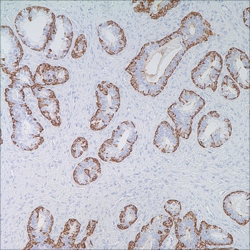
Calretinin (SP13) Rabbit Monoclonal Antibody

Sign Into View Organizational & Contract Pricing


About This Item

UNSPSC Code:
12352203
NACRES:
NA.41

biological source

rabbit

Quality Level

100
500

conjugate

unconjugated

antibody form

culture supernatant

antibody product type

primary antibodies

clone

SP13, monoclonal

description

For In Vitro Diagnostic Use in Select Regions (See Chart)

form

buffered aqueous solution

species reactivity

human

packaging

vial of 0.1 mL concentrate (232R-14)
vial of 0.5 mL concentrate (232R-15)
bottle of 1.0 mL predilute (232R-17)
vial of 1.0 mL concentrate (232R-16)
bottle of 7.0 mL predilute (232R-18)

manufacturer/tradename

Cell Marque

technique(s)

immunohistochemistry (formalin-fixed, paraffin-embedded sections): 1:25-1:100

isotype

IgG1

control

mesothelioma

shipped in

wet ice

storage temp.

2-8°C

visualization

cytoplasmic, nuclear

General description

Calretinin is a 29 kDa calcium-binding protein thought to play a role in the cell cycle. Anti-calretinin has been shown to be useful in differentiating mesothelioma from adenocarcinomas from the lung and other sources. The sensitivity of anti-calretinin in distinguishing reactive mesothelial cells from adenocarcinoma cells is 100%, and the specificity is up to 80%. Light, focal, cytoplasmic staining of adenocarcinoma cells may be seen in 10 to 30% of cases. Anti-calretinin has also demonstrated utility in differentiating adrenal cortical neoplasms (+) from pheochromocytomas (-). Calretinin has also been useful in diagnosing granulosa cell tumor of the ovary amongst its mimics. Other ovarian tumors that stain with calretinin are Sertoli-Leydig cell tumor, Sertoli cell tumor, Leydig cell tumor, and sex cord tumor with annular tubules, as well as steroid cell tumor.9

Linkage

Calretinin Positive Control Slides, Product No. 232S, are available for immunohistochemistry (formalin-fixed, paraffin-embedded sections).

Physical form

Solution in Tris Buffer, pH 7.3-7.7, with 1% BSA and <0.1% Sodium Azide

Preparation Note

Download the IFU specific to your product lot and formatNote: This requires a keycode which can be found on your packaging or product label.

Other Notes

For Technical Service please contact: 800-665-7284 or email: service@cellmarque.com

Legal Information

Cell Marque is a trademark of Merck KGaA, Darmstadt, Germany

Not finding the right product?  

Try our Product Selector Tool.

Storage Class Code

12 - Non Combustible Liquids

WGK

WGK 2

Flash Point(F)

Not applicable

Flash Point(C)

Not applicable


Choose from one of the most recent versions:

Certificates of Analysis (COA)

Lot/Batch Number

Don't see the Right Version?

If you require a particular version, you can look up a specific certificate by the Lot or Batch number.

Already Own This Product?

Find documentation for the products that you have recently purchased in the Document Library.

Visit the Document Library

Paul J Zhang et al.
Modern pathology : an official journal of the United States and Canadian Academy of Pathology, Inc, 16(6), 591-597 (2003-06-17)
Morphologic distinction between adrenal cortical and medullary tumors can be difficult. Previous studies have shown inhibin, melan-A, and BCL-2 to be useful markers for adrenal cortical tumors. We have recently observed a high level of calretinin expression in normal adrenal
M J Costa et al.
Human pathology, 28(11), 1247-1254 (1997-12-31)
Immunohistochemistry using monoclonal antibodies against human inhibin, a peptide hormone produced by ovarian granulosa cells to inhibit follicle-stimulating hormone (FSH), has been recently applied to diagnostic anatomic pathology. This investigation hypothesizes that inhibin immunohistochemistry will aid in the crucial clinical
M Rishi et al.
The American journal of surgical pathology, 21(5), 583-589 (1997-05-01)
Inhibin is a glycoprotein hormone produced by normal ovarian granulosa cells and testicular sertoli cells. In the ovary, it inhibits the secretion of follicle-stimulating hormone. Patients with granulosa cell tumors (GCT) have elevated serum levels of inhibin and this finding
Merce Jorda et al.
Applied immunohistochemistry & molecular morphology : AIMM, 10(1), 67-70 (2002-03-15)
Most adrenocortical neoplasms and pheochromocytomas can be diagnosed by a combination of clinical findings and morphologic features. Occasionally, however, this histologic differential diagnosis requires ancillary tests, such as immunohistochemistry. Both tumors are generally negative for epithelial markers but express synaptophysin.
Wenxin Zheng et al.
Advances in anatomic pathology, 10(1), 27-38 (2002-12-28)
Through a brief introduction of inhibin history, characteristics of the antibody against inhibin, and normal tissue distribution of alpha-inhibin expression, this comprehensive review focuses on a practical approach to using alpha-inhibin in the differential diagnosis of ovarian sex cord-stromal tumors

Our team of scientists has experience in all areas of research including Life Science, Material Science, Chemical Synthesis, Chromatography, Analytical and many others.

Contact Technical Service