11333062910
Roche
Anti-Digoxigenin
from mouse IgG1κ (clone 1.71.256)
Synonym(s):
anti-digoxigenin, digoxigenin
Sign Into View Organizational & Contract Pricing
All Photos(1)
About This Item
UNSPSC Code:
12352203
Recommended Products
biological source
mouse
Quality Level
conjugate
unconjugated
antibody form
purified immunoglobulin
antibody product type
primary antibodies
clone
clone 1.71.256, monoclonal
form
lyophilized
packaging
pkg of 100 μg
manufacturer/tradename
Roche
technique(s)
ELISA: 2-4 μg/mL
immunocytochemistry: 0.5-2 μg/mL
immunohistochemistry: 0.5-2 μg/mL
western blot: 0.5-2 μg/mL
isotype
IgG1κ
storage temp.
2-8°C
General description
Contents: Lyophilizate
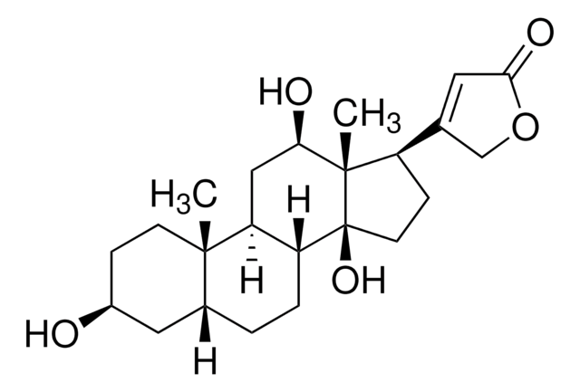
Digoxigenin is a hapten, useful in labeling nucleic acids and in detection systems. Probes labeled with digoxigenin has greater sensitivity equivalent to that of radioactive probes, allows faster detection. It is less hazardous and has increased shelf life.
Specificity
The monoclonal antibody reacts with free and bound digoxigenin.
Application
Use Anti-Digoxigenin antibody for the detection of digoxigenin-labeled componds using:
- ELISA
- Immunohistocytochemistry
- In situ hybridization
- Western blot
- Immunofluorescence staining
- FISH (fluorescent in situ hybridization)
Biochem/physiol Actions
In the presence of Na+, Mg2+ and adenosine triphosphate (ATP), digoxigenin inhibits sodium pumps.
Features and Benefits
- Specific to free and bound digoxigenin
- The antibody is not stabilized with protein and therefore suitable as coating antibody in Sandwich-ELISAs and for labeling procedures. For example it can be conjugated with enzymes or fluorescence dyes for direct detection of digoxigenin-labeled samples.
Preparation Note
Working concentration: Working concentration of antibody depends on application and substrate. The following concentrations should be taken as a guideline:
Working solution: For coating applications
phosphate buffered saline, pH 7.4
- ELISA: for coating: 2 to 4 μg/ml
- Immunohistocytochemistry: 0.5 to 2 μg/ml
- In situ hybridization: 0.2 to 0.4 μg/ml
- Western blot: 0.5 to 2 μg/ml
Working solution: For coating applications
phosphate buffered saline, pH 7.4
Reconstitution
Add 1 ml double-distilled water to a final concentration of 100 μg/ml.
Storage and Stability
The lyophilized antibody is stable at +2 to +8°C. The reconstituted antibody solution is stable up to 6 months at +2 to +8°C. The solution can be stored in aliquots at -15 to -25°C. Avoid repeated freezing and thawing.
Analysis Note
There are no cross reactivities known.
Other Notes
For life science research only. Not for use in diagnostic procedures.
Not finding the right product?
Try our Product Selector Tool.
Signal Word
Warning
Hazard Statements
Precautionary Statements
Hazard Classifications
Skin Sens. 1
Storage Class Code
11 - Combustible Solids
WGK
WGK 3
Flash Point(F)
does not flash
Flash Point(C)
does not flash
Choose from one of the most recent versions:
Already Own This Product?
Find documentation for the products that you have recently purchased in the Document Library.
Customers Also Viewed
Xingqi Chen et al.
BioTechniques, 56(3), 117-118 (2014-03-20)
Current techniques for analyzing chromatin structures are hampered by either poor resolution at the individual cell level or the need for a large number of cells to obtain higher resolution. This is a major problem as it hampers our understanding
Affinity Labeling of a Sulfhydryl Group in the Cardiacglycoside Receptor Site of Na+/K+-ATPase by N-Hydroxysuccinimidyl Derivatives of Digoxigenin
<BIG>Antolovic R, et al.</BIG>
European Journal of Biochemistry, 227, 61-67 (1995)
Carla Winterling et al.
RNA biology, 11(1), 66-75 (2014-01-21)
A growing body of evidence suggests the non-protein coding human genome is of vital importance for human cell function. Besides small RNAs, the diverse class of long non-coding RNAs (lncRNAs) recently came into focus. However, their relevance for infection, a
Nonradioactive labeling of probe with digoxigenin by polymerase chain reaction
<BIG>Lion T and Haas OA</BIG>
Analytical Chemistry, 188, 335-337 (1990)
Kristiina Joensuu et al.
Breast cancer : basic and clinical research, 7, 23-34 (2013-03-22)
Breast cancer can recur even decades after the primary therapy. Markers are needed to predict cancer progression and the risk of late recurrence. The estrogen receptor (ER), progesterone receptor (PR), human epidermal growth factor receptor-2 (HER2), proliferation marker Ki-67, and
Our team of scientists has experience in all areas of research including Life Science, Material Science, Chemical Synthesis, Chromatography, Analytical and many others.
Contact Technical Service


