Skip to Content
Merck
All Photos(1)

Key Documents

AB3639

Sigma-Aldrich

Anti-Endonuclease G Antibody

Chemicon®, from rabbit

Synonym(s):

EndoG

Sign Into View Organizational & Contract Pricing


About This Item

UNSPSC Code:
12352203
eCl@ss:
32160702
NACRES:
NA.41

biological source

rabbit

Quality Level

antibody form

affinity purified immunoglobulin

antibody product type

primary antibodies

clone

polyclonal

purified by

affinity chromatography

species reactivity

mouse, rat, human

manufacturer/tradename

Chemicon®

technique(s)

western blot: suitable

NCBI accession no.

UniProt accession no.

shipped in

wet ice

target post-translational modification

unmodified

Gene Information

human ... ENDOG(2021)
mouse ... Endog(13804)
rat ... Endog(362100)

Specificity

Recognizes Endonuclease G (EndoG), a mitochondrion-specific nuclease that translocates to the nucleus and cleaves chromatin DNA during apoptosis. EndoG is specifically activated by apoptotic stimuli and is able to induce nucleosomal fragmentation of DNA independently of caspase and DFF/CAD (1,2).

Immunogen

Synthetic peptide corresponding to amino acids 55 to 70 of human EndoG (GGPRGPGELAKYGLP).

Application

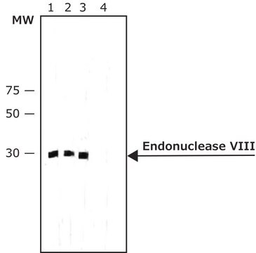
Anti-Endonuclease G Antibody is an antibody against Endonuclease G for use in WB.
Research Category
Apoptosis & Cancer
Research Sub Category
Apoptosis - Additional
Western blotting (1-2μg/mL)

Optimal working dilutions must be determined by the end user.

Physical form

Affinity purified immunoglobulin. Liquid in PBS containing 0.02% sodium azide.

Storage and Stability

Maintain at 2-8ºC for 12 months.

Other Notes

Concentration: Please refer to the Certificate of Analysis for the lot-specific concentration.

Legal Information

CHEMICON is a registered trademark of Merck KGaA, Darmstadt, Germany

Disclaimer

Unless otherwise stated in our catalog or other company documentation accompanying the product(s), our products are intended for research use only and are not to be used for any other purpose, which includes but is not limited to, unauthorized commercial uses, in vitro diagnostic uses, ex vivo or in vivo therapeutic uses or any type of consumption or application to humans or animals.

Not finding the right product?  

Try our Product Selector Tool.

Storage Class Code

10 - Combustible liquids

WGK

WGK 2

Flash Point(F)

Not applicable

Flash Point(C)

Not applicable


Certificates of Analysis (COA)

Search for Certificates of Analysis (COA) by entering the products Lot/Batch Number. Lot and Batch Numbers can be found on a product’s label following the words ‘Lot’ or ‘Batch’.

Already Own This Product?

Find documentation for the products that you have recently purchased in the Document Library.

Visit the Document Library

Tamara Azarashvili et al.
International journal of molecular sciences, 17(12) (2016-12-17)
The translocator protein (TSPO; 18 kDa) is a high-affinity cholesterol-binding protein located in the outer membrane of mitochondria. A domain in the C-terminus of TSPO was characterized as the cholesterol recognition/interaction amino acid consensus (CRAC). The ability of the CRAC
Shuangping Zhao et al.
Journal of neuroscience research, 88(8), 1727-1737 (2010-01-16)
We have shown that generalized seizures produce necrotic neurons with caspase-independent nuclear pyknosis and DNA fragmentation. In this study, we determined the time course of translocation of mitochondrial cytochrome c, apoptosis-inducing factor, endonuclease G, lysosomal cathepsins B and D, and
H-W Zheng et al.
Cell death & disease, 5, e1262-e1262 (2014-05-31)
Receptor-interacting protein (RIP) kinases promote the induction of necrotic cell death pathways. Here we investigated signaling pathways in outer hair cells (OHCs) of adult male CBA/J mice exposed to noise that causes permanent threshold shifts, with a particular focus on
Traumatic noise activates Rho-family GTPases through transient cellular energy depletion.
Chen, FQ; Zheng, HW; Hill, K; Sha, SH
The Journal of Neuroscience null
Sathivel Chinnathambi et al.
Cells, tissues, organs, 187(2), 131-140 (2007-10-17)
We examined how young and old keratinocytes died from heat stress in vitro. We found that keratinocyte cell death was not due to oxidative stress as neither Mn-SOD nor Cu-Zn-SOD was produced in either young or old heated keratinocytes. Instead

Our team of scientists has experience in all areas of research including Life Science, Material Science, Chemical Synthesis, Chromatography, Analytical and many others.

Contact Technical Service