Saltar al contenido
Merck
Todas las fotos(3)

Documentos

MABN783

Sigma-Aldrich

Anti-Semaphorin 3A Antibody, clone Sema-3A

clone Sema-3A, from mouse

Sinónimos:

Collapsin-1, COLL-1, Sema-3A

Iniciar sesiónpara Ver la Fijación de precios por contrato y de la organización


About This Item

UNSPSC Code:
12352203
eCl@ss:
32160702
NACRES:
NA.41

biological source

mouse

Quality Level

antibody form

purified immunoglobulin

antibody product type

primary antibodies

clone

Sema-3A, monoclonal

species reactivity

rabbit, chicken, human, rat, fish, mouse

technique(s)

immunofluorescence: suitable
immunohistochemistry: suitable (paraffin)
western blot: suitable

isotype

IgG1κ

NCBI accession no.

UniProt accession no.

shipped in

ambient

target post-translational modification

unmodified

Gene Information

human ... SEMA3A(10371)

General description

Semaphorin-3A (UniProt: Q90607, also known as Collapsin-1, COLL-1) is encoded by the SEMA3A (also known as COLL1) gene (gene ID: 395825) in chicken. Semaphorin-3A is a secreted protein that is synthesized with a 22 amino acid signal peptide. It is a member of a family of proteins that are involved in cell migration and axonal growth cone guidance. Semaphorin-3A has an Ig-like C2-type (immunoglobulin-like) domain, a PSI domain, and a Sema domain of 500 amino acids. It strongly binds to neuropilin via its carboxy third sequence and competes with semaphorin dimerization. It is expressed at relatively high levels in brain and muscle and at moderate levels in lung, bursa, and heart. It is not found in the liver. Semaphorin-3A can function as either a chemorepulsive agent, inhibiting axonal outgrowth, or as a chemoattractive agent, stimulating the growth of apical dendrites. It induces the collapse and paralysis of neuronal growth cones and could serve as a ligand that guides specific growth cones by a motility-inhibiting mechanism. Increased expression of Semaphorin-3A is associated with schizophrenia and it is seen in a variety of human tumor cell lines. Additionally, aberrant release of this protein is associated with the progression of Alzheimer′s disease. Semaphorin-3A is also reported to exert an osteoprotective effect by both suppressing osteoclastic bone resorption and increasing osteoblastic bone formation. The downregulation of Semaphorin-3A expression is shown to cause ischemia, degenerative diseases, and multiple sclerosis. (Ref.: Hayashi, M et al. (2012). Nature. 485(7396):69-74).

Specificity

Clone Sema-3A specifically targets Semaphorin 3A in multiple species. It targets a sequence of 18 amino acids within the N-terminal half of Semaphorin 3A.

Immunogen

Epitope: unknown
KLH-conjugated linear peptide corresponding to 18 amino acids from the N-terminal half of Chicken Semaphorin-3A.

Application

Anti-Semaphorin 3A Antibody, clone Sema-3A, Cat. No. MABN783, is a highly specific mouse monoclonal antibody that targets Semaphorin 3A and has been tested in Immunofluorescence, Immunohistochemistry (Paraffin), and Western Blotting.
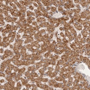
Immunohistochemistry Analysis: A 1:5,000 dilution from a representative lot detected Semaphorin 3A in human cerebellum, human cerebral cortex, rat cerebral cortex, and rat kidney tissues.

Immunohistochemistry Analysis: A representative lot detected Semaphorin 3A in Immunohistochemistry applications (Rosenzweig, S., et. al. (2010). Graefes Arch Clin Exp Opthalmol. 248(10):1423-35).

Immunofluorescence Analysis: A representative lot detected Semaphorin 3A in Immunofluorescence applications (Nitzan, A., et. al. (2006). Glia. 54(6):545-56; Baranes, K., et. al. (2009). Exp Neurol. 218(1):24-32).

Western Blotting Analysis: A representative lot detected Semaphorin 3A in Western Blotting applications (Nitzan, A., et. al. (2006). Glia. 54(6):545-56; Rosenzweig, S., et. al. (2010). Graefes Arch Clin Exp Opthalmol. 248(10):1423-35).
Research Category
Neuroscience

Quality

Evaluated by Western Blotting in HEK293 cell culture enriched with Semaphorin 3A.

Western Blotting Analysis: 0.01 µg/mL of this antibody detected Semaphorin 3A in 23 µL of enriched culture supernatant from HEK293 cells.

Target description

~95 kDa observed; 88.87 kDa calculated. Uncharacterized bands may be observed in some lysate(s).

Physical form

Format: Purified
Protein G purified
Purified mouse monoclonal antibody IgG1 in buffer containing 0.1 M Tris-Glycine (pH 7.4), 150 mM NaCl with 0.05% sodium azide.

Storage and Stability

Stable for 1 year at 2-8°C from date of receipt.

Other Notes

Concentration: Please refer to lot specific datasheet.

Disclaimer

Unless otherwise stated in our catalog or other company documentation accompanying the product(s), our products are intended for research use only and are not to be used for any other purpose, which includes but is not limited to, unauthorized commercial uses, in vitro diagnostic uses, ex vivo or in vivo therapeutic uses or any type of consumption or application to humans or animals.

¿No encuentra el producto adecuado?  

Pruebe nuestro Herramienta de selección de productos.

Storage Class

12 - Non Combustible Liquids

wgk_germany

WGK 1


Certificados de análisis (COA)

Busque Certificados de análisis (COA) introduciendo el número de lote del producto. Los números de lote se encuentran en la etiqueta del producto después de las palabras «Lot» o «Batch»

¿Ya tiene este producto?

Encuentre la documentación para los productos que ha comprado recientemente en la Biblioteca de documentos.

Visite la Librería de documentos

Anat Nitzan et al.
Translational vision science & technology, 10(10), 16-16 (2021-11-25)
Semaphorin 3A (Sema-3A) is a secreted protein that deflects axons from inappropriate regions and induces neuronal cell death. Intravitreal application of polyclonal antibodies against Sema-3A prevents loss of retinal ganglion cells ensuing from axotomy of optic nerves. This suggested a

Nuestro equipo de científicos tiene experiencia en todas las áreas de investigación: Ciencias de la vida, Ciencia de los materiales, Síntesis química, Cromatografía, Analítica y muchas otras.

Póngase en contacto con el Servicio técnico